Age: 45Yrs
Sex: Female
Case study: Uterus: Normal in size and shows two separate uteri with widely divergent apices, two separate cervices noted. Normal uterine zone anatomy is preserved in each uteri. Mildly heterogeneous myometrium noted in the right uteri with junctional zone measuring 10 mm. Intramural fibroid measuring 10 x 9 mm noted in the medial wall of left uteri.
Images:
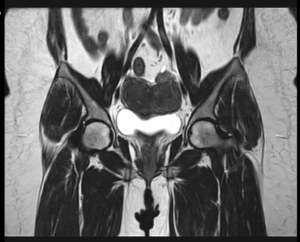
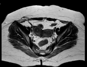
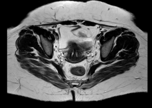
Conclusion : Imaging features are suggestive of uterine didelphys with intramural fibroid in left uteri and heterogeneous myometrium of right uteri (? Adenomyosis) as described.