Age: 11 Yrs
Sex: Female
Complaints: Developmental abnormalities. K/c/o HIE.
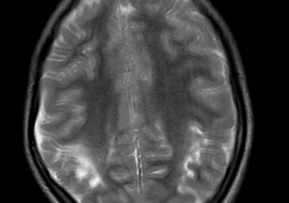
Case study: Evidence of periventricular T2/FLAIR hyperintensities seen in the posterior fossa around the occipital horn extending into the adjacent sulcal spaces. The corpus callosum shows significant thinning predominantly involving the isthmus and splenium. Volume loss seen in the right hippocampal head with widening of CSF space. There is asymmetric ex-vacuo dilatation of right occipital horn of lateral ventricle.
Images:
![]()

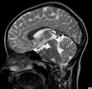

Conclusion: Atrophic changes involving the bilateral cerebral hemisphere in the form of periventricular and deep white matter hyperintensities showing cortical thinning along with asymmetric ex-vacuo dilatation of occipital horn. Corpus callosal thinning involving the isthmus and splenium. Right hippocampal volume loss. – Features are in consistent with atrophic changes as sequelae of Hypoxic Ischemic Encephalopathy.