Head
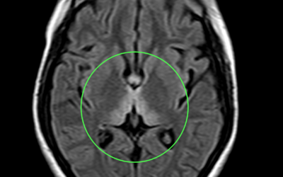
Wernicke Encephalopathy
Age: 27yr Sex: Female Complaints: Gait ataxia and ophthalmoplegia confusion. Case study: Areas of symmetrical increased T2/FLAIR signal seen involving dorsomedial thalami, tectal plate, periaqueductal area, around 3rd ventricle, mammillary bodies, posterior medulla...
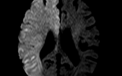
Acute Cerebral Infarct
Age: 74yr Sex: Female Complaints: Left limb weakness. Case study: Large area of diffusion restriction with corresponding low ADC signal seen involving right fronto- parieto- temporal lobe with no signal abnormalities in corresponding T2 and FLAIR sequence.Angiography...
Acute Pontine Hemorrhage
Age: 68 Yrs Sex: Female Complaints: Spontaneous headache. Case study: A well defined hyperdense (max. ~60HU) area measuring ~19 x 16 x 6 mm (TR x CC x AP) noted in the left pontine hemisphere extending – Features in favor of acute pontine hemorrhage. No...
Acute on chronic SDH
Name: Aliyaru kunju Age: 62 Yrs Sex: Male Complaints: c/o ataxia Introduction: Case study: Bilateral cerebral hemisphere shows well defined encapsulated hypodense subdural collection involving right parietal and left fronto-parietal region with maximum thickness in...
Acute on chronic infarct
Name: Ramachandran Age: 73 Yrs Sex: Male Complaints: H/o fall. Headache Introduction: Case study: Bilateral cerebral hemisphere shows well defined irregular hypodense (16-20HU) encapsulated subdural collection with maximum thickness measuring ~7 mm in the left high...
Acute on chronic SDH
Case: 23 Heading: Acute on chronic SDH Name: Dr. Ahammed Kunju Age: 79 Yrs Sex: Male Complaints: H/o fall. Introduction: Case study: Bilateral cerebral hemisphere shows well defined encapsulated hypodense subdural collection with maximum thickness in the right...
Inter-hemispheric bleed
Name: Sudevan Age: 54 Sex: Male Complaints: Introduction: H/o trauma Case study: Thin linear hyperdensity noted involving the posterior inter-hemispheric fissure and right tentorium cerebelli. – Suggestive of acute subdural hemorrhage (SDH). Images Conclusion: Acute...
Watershed Infarcts
Age: 61yr Sex: Female Complaints: H/o deviation of angle of mouth Case study: Poorly defined scattered irregular hypodensities (max. 18HU) noted in the bilateral high parietal region involving the corona radiata and centrum semi ovale. – Features suggestive of...
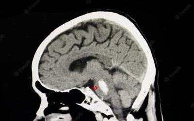
Craniopharyngioma
Name: Ambili Age: 47 Sex: Female Complaints: H/o left vision deficit. Introduction: Case study: A well defined solid lesion with peripheral hyperdensity in the sellar region measuring ~14 x 13 mm. Sagittally the lesion is seen extending into the pituitary fossa....
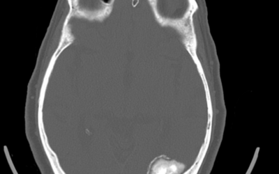
A case of atypical meningioma
CALCIFIED MENINGIOMA A 52yr old woman with intermittent head ache and syncope. Non Contrast Computed Tomography[NCCT] shows a well-defined irregular space occupying calcified lesion in the left occipital lobe.


