Sex: Male
Complaints: Left Flank pain.
Age: 48yr
Case study: Left kidney: 10.8 X 3.6 cm. Minimal perinephric free fluid with fat stranding noted. A calculus (~HU 581) measuring ~ 7.7 x 5.7 x 5.4 mm (CC x AP x TR)noted in the upper segment of left ureter with periureteric fat stranding through out its course.
Images:
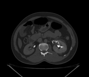


On contrast administration left kidney in delayed phase:
4 min delay image shows : No contrast transport in the ureter.
10 min delay image shows: No contrast transport in the ureter. There is evidence of contrast extravasation tracing from the renal hila extending circumferentially around proximal ureter confined to the upper segment.
20 min delay image shows: No contrast transport in the ureter. There is evidence of contrast extravasation tracing from the renal hila and mid calyx extending into the perinephric and periureteric region. There is irregular streaking of contrast extending into the adjacent perinephric plane both medially and laterally.
30 min delay images shows: No contrast transport in the left ureter. There is further more extravasation of contrast along the ureter caudally in addition to previously seen mentioned areas.
Conclusion: Left ureteric calculus with renal inflammatory changes in the form of perinephric fluid and fat stranding. Left kidney shows contrast leakage tracing from the hila as described in delayed phase with increase in area of spread proportionate to increase in time. – ?pelvic/calyceal rupture. Left ureter shows no contrast transport through out the multiple phase imaging study. No urinary bladder calculus.