Age: 48 Yrs
Sex: Female
Complaints: H/o left chest wall swelling. No pain or skin abnormality.
Images:
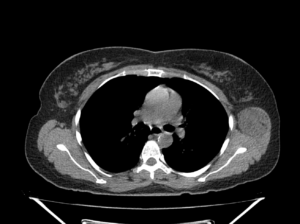
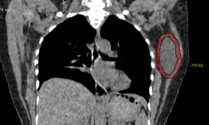
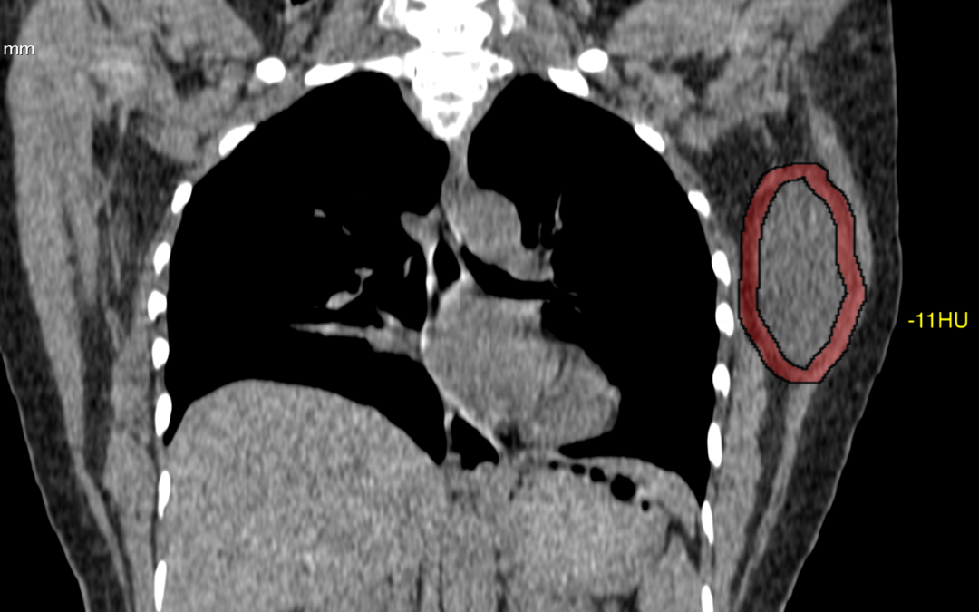
Well circumscribed hypo to isodense (max. -15HU) oval shaped mass lesion measuring ~6.6 x 4.0 x 3.8 cm (CC x TR x AP) noted evolving from the left latissimus dorsi and muscle involving the intramuscular space between subscapularis. No evidence of any bone involvement.
Case Discussion :
Well defined smooth solid hypo to iso-dense lesion in the left chest wall seen arising from the latissimus dorsi muscle.
The is lesion is seen as a solid mass with hypo-dense attenuation, however the lesion shows typical fatty appearance.
The report was given as chest wall mass arising from left latissimus dorsi muscle.
Possible DD’s include: 1. Lipoma. 2. Liposarcoma 3.Schwannoma.
Conclusion: FNA done. Histopathology came as fat containing lesion and suggested Excision biopsy.
Reference
- Lee JH, Do HD, Lee JC. Well-circumscribed type of intramuscular lipoma in the chest wall. Journal of cardiothoracic surgery. 8: 181. doi:10.1186/1749-8090-8-181 – Pubmed
- 3. López Soriano A, Tomasello A, Luburich P, Noel A. Fat necrosis in a chest wall lipoma. AJR. American journal of roentgenology. 183 (3): 866. doi:10.2214/ajr.183.3.1830866 – Pubmed