by Nithesh Ravindran | Dec 15, 2021
Age: 48 Yrs
Sex: Female
Complaints: H/o left chest wall swelling. No pain or skin abnormality.
Images:
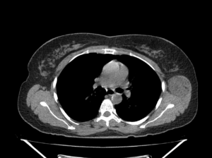
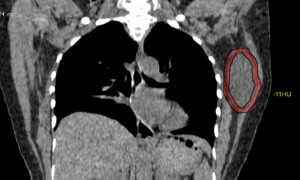
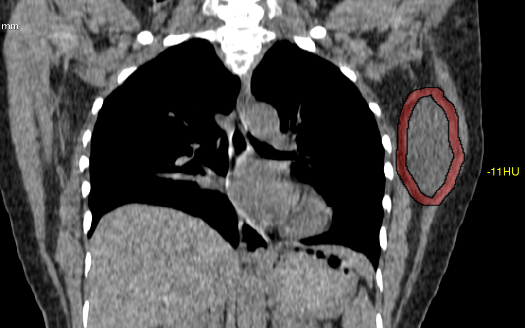
Well circumscribed hypo to isodense (max. -15HU) oval shaped mass lesion measuring ~6.6 x 4.0 x 3.8 cm (CC x TR x AP) noted evolving from the left latissimus dorsi and muscle involving the intramuscular space between subscapularis. No evidence of any bone involvement.
Case Discussion :
Well defined smooth solid hypo to iso-dense lesion in the left chest wall seen arising from the latissimus dorsi muscle.
The is lesion is seen as a solid mass with hypo-dense attenuation, however the lesion shows typical fatty appearance.
The report was given as chest wall mass arising from left latissimus dorsi muscle.
Possible DD’s include: 1. Lipoma. 2. Liposarcoma 3.Schwannoma.
Conclusion: FNA done. Histopathology came as fat containing lesion and suggested Excision biopsy.
Reference
- Lee JH, Do HD, Lee JC. Well-circumscribed type of intramuscular lipoma in the chest wall. Journal of cardiothoracic surgery. 8: 181. doi:10.1186/1749-8090-8-181 – Pubmed
- 3. López Soriano A, Tomasello A, Luburich P, Noel A. Fat necrosis in a chest wall lipoma. AJR. American journal of roentgenology. 183 (3): 866. doi:10.2214/ajr.183.3.1830866 – Pubmed

by Nithesh Ravindran | Oct 27, 2021
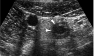
Transitional cell carcinoma (TCC), also called urothelial cell carcinoma (UCC) of the bladder, is the most common primary neoplasm of the urinary bladder, and bladder TCC is the most common tumour of the entire urinary system.
CASE DISCUSSION
The bladder is by far the most common site of transitional cell carcinomas, 50 times more common than TCC of the renal pelvis, and 100 times more common than TCC of the ureter 1. Bladder TCCs are the most common tumour of the entire urinary tract.
There is a known association of TCCs developing within bladder diverticula, presumably due to urinary stasis which leads to chronic urothelial irritation and potentially exaggerated exposure to urinary carcinogens 10-12.
by Nithesh Ravindran | Oct 26, 2021
Age: 52 Yrs
Sex: Female
Complaints: A 52yr old male came with chief complaints of abdominal distension with increase tension in the abdomen during standing.
Images:


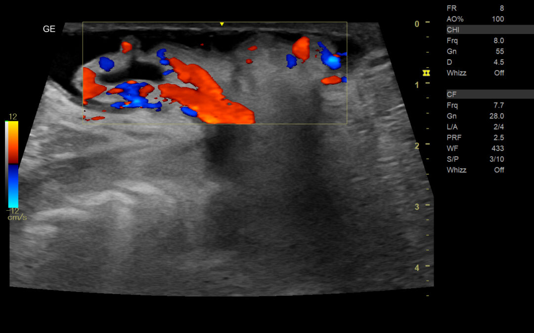
On color doppler evaluation there is evidence of mesenteric vessel showing significant vascularity along with the herniation of fat.
Ultrasound images shows an abdominal wall defect in the umbilical region with herniation omental and evidence free fluid in the herniated sac.

by admin | Oct 20, 2021
Age: 13yr
Sex: Female
Complaints: Right lower abdominal pain.
Case study: RIF: Ultrasound examination shows enlarged appendix measuring ~8.7mm with non-compressible oedematous wall and minimal peri-appendiceal fluid.
Images:
A case of acute appendicitis.Ultrasound examination shows enlarged appendix measuring ~8.7mm with non-compressible oedematous wall and minimal peri-appendiceal fluid.
by admin | Oct 20, 2021
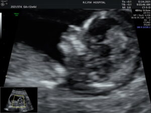
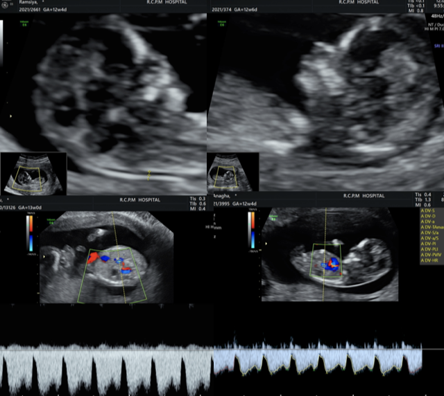
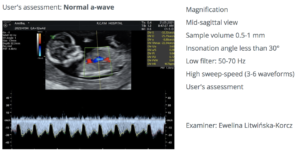
FMF CERTIFIED IMAGE OF NASAL BONE IN MID SAGITTAL SECTION.
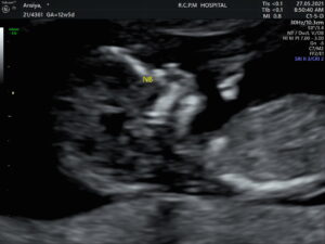
ANOTHER IMAGE SHOWING NASAL TIP, SKIN AND NASAL BONE
![FMF CERTIFIED NUCHAL TRANSLUCENY [NT] IN TRUE MID SAGITTAL SECTION](http://nrradiology.com/wp-content/uploads/2021/10/IMG_20210413_6_1-300x225.jpg)
NUCHAL TRANSLUCENCY IN MID SAGGITTAL VIEW

by admin | Oct 20, 2021
CALCIFIED MENINGIOMA
A 52yr old woman with intermittent head ache and syncope. Non Contrast Computed Tomography[NCCT] shows a well-defined irregular space occupying calcified lesion in the left occipital lobe.
