
by Nithesh Ravindran | Dec 5, 2022
Case Courtesy : Dr Sajith Selvaganesan MD FSCCT FSCMR,Sunrise Hospital
Age: 25Yrs
Sex: Male
Complaints: Lower back pain.
Case study:
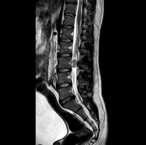
Loss of normal lumbar lordosis.
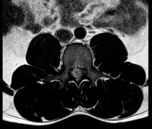
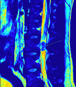
Relatively well defined, mildly heterogeneous, round to oval, iso to hyperintense intramedullary lesion measuring 11 x 10 x 17 mm (TR x AP x CC) noted the level of L3 vertebra
Images:
Conclusion
Relatively well defined, mildly heterogeneous, round to oval, iso to hyperintense intramedullary lesion measuring 11 x 10 x 17 mm (TR x AP x CC) noted the level of L3 vertebra – Likely myxopapillary ependymoma. Differential includes paraganglioma. Suggested HPE evaluation.
by Nithesh Ravindran | Dec 4, 2022
Case courtesy : Dr Sajith Selvaganesan MD FSCCT FSCMR, Sunrise Hospital
Age: 68 Yrs
Sex: Female
Case study:
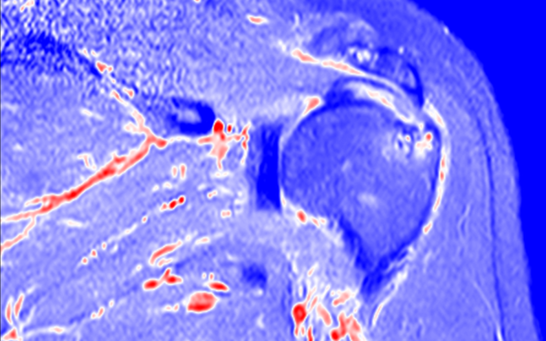
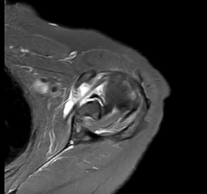
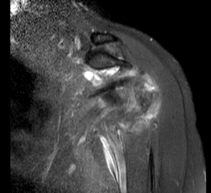
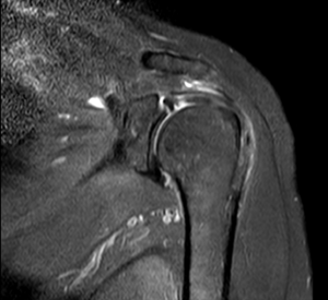
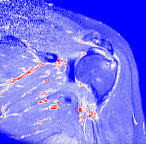
High grade partial tear of subscapularis tendon with no retraction of muscle.
Altered intra-substance signal intensity noted in the supraspinatus tendon reaching up to the articular surface.
Altered signal intensity noted in the posterolateral aspect of humeral head.
Type V superior labral anterior posterior tear noted with minimal fluid along the interface.
Type II acromion seen.
Minimal joint effusion with subcoracoid bursal fluid.
Images:
Conclusion :
High grade partial tear of subscapularis tendon.
Partial tear of supraspinatus tendon.
Type V superior labral anterior posterior tear.
Altered signal intensity in the posterolateral aspect of humeral head – Likely hillsach’s lesion.
Minimal joint effusion with subcoracoid bursal fluid.
Acromioclavicular joint athropathy changes.
by Nithesh Ravindran | Oct 11, 2022
Age: 30Yrs
Sex: Male
Complaints: Pain on foot while walking.
Case study: Subchondral high signal noted in the mid aspect of the talus and the calcaneum with small subchondral cystic areas – Apparent on the STIR sequences. Evidence of osteophytic changes with cortical irregularity and narrowing of the talo-calcaneal joint anteriorly. The osteophyte is seen encroaching into the sinus tarsi with narrowing.
The tibio-calcaneal angle (kites angle) is 83 degrees – Normal.
The talo-calcaneal angle is 45 degrees – Abnormal.
Images:
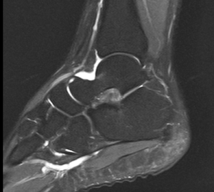
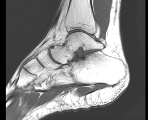
Conclusion : Subchondral talo-calcaneal high marrow signals with anterior joint space narrowing, osteophytic formation and encroachment into the sinus tarsi – Suggestive of talo-calcaneal impingement.
Increased talo-calcaneal angle (45 degrees) – Hindfoot valgus.
Peroneus longus and brevis tenosynovitis.
by Nithesh Ravindran | Dec 9, 2021
Name: Jaleela Ahammed
Age: 59 Yrs
Sex: Female
Complaints: To r/o abscess.
Introduction:
Case study: Well defined thick walled heterogenous appearing hypoechoic collection measuring ~24 x 12 mm noted in the subcutaneous region of left lower abdomen. No evidence of any internal vascularity.
Images
Conclusion: Well defined thick walled hypoechoic collection in the subcutaneous plane of left lower abdomen at the site of needle puncture. – Features in favour of post injection abscess.
Reference :