by Nithesh Ravindran | Jan 27, 2022
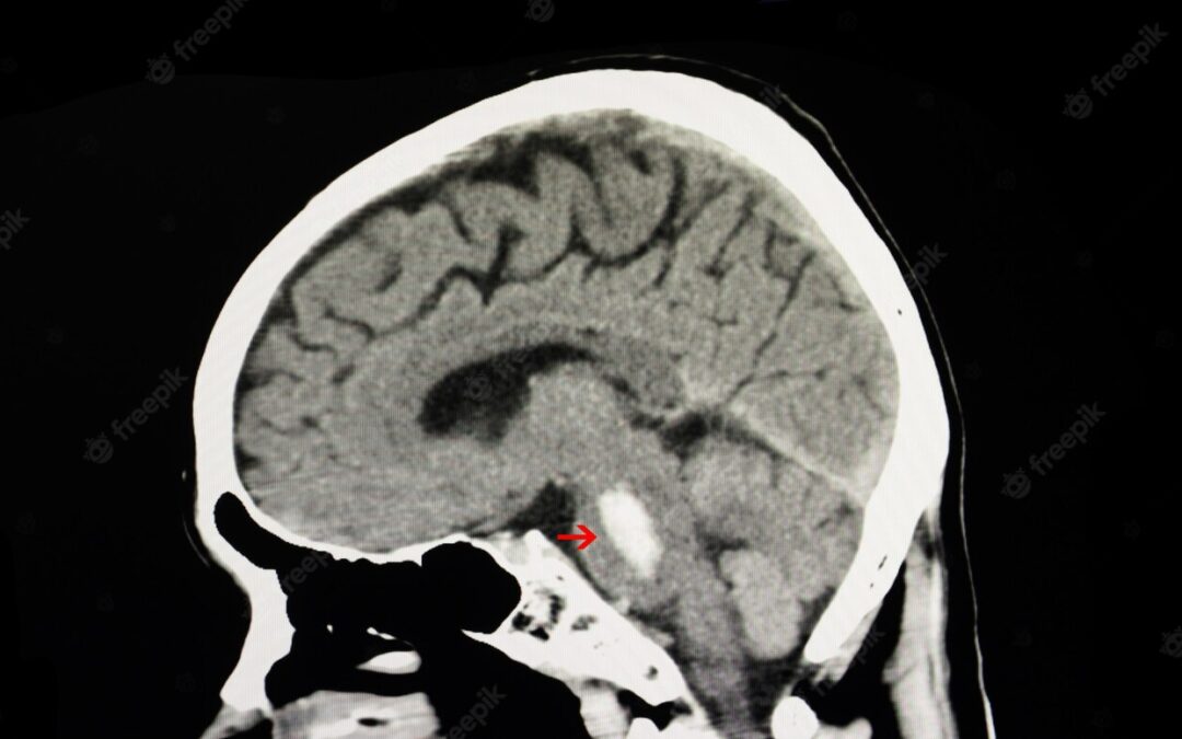
Age: 68 Yrs
Sex: Female
Complaints: Spontaneous headache.
Case study: A well defined hyperdense (max. ~60HU) area measuring ~19 x 16 x 6 mm (TR x CC x AP) noted in the left pontine hemisphere extending – Features in favor of acute pontine hemorrhage. No evidence of mass effect/hydrocephalus/intra-ventricular hemorrhage.
Conclusion: Acute pontine hemorrhage . – Features in favor of hypertensive etiology.
by Nithesh Ravindran | Dec 30, 2021
Name: Aliyaru kunju
Age: 62 Yrs
Sex: Male
Complaints: c/o ataxia
Introduction:
- Case study: Bilateral cerebral hemisphere shows well defined encapsulated hypodense subdural collection involving right parietal and left fronto-parietal region with maximum thickness in the right parietal region measuring ~28 mm showing irregular areas of hyperdensities.
- Well defined encapsulated hypodense collection measuring ~4.7 x 1.5 cm involving the epidural space of right frontal lobe extending into the parietal region with minimal evidence of peripheral hyperdensities.
- No evidence of significant mass effect/ midline shift at present.
Images
Conclusion: Well defined encapsulated subdural hypodense collections in the bilateral cerebral hemisphere with evidence of hyperdensities. – Features in favor of acute on chronic SDH.
Well defined encapsulated epidural hypodense collection in the right fronto-parietal region with peripheral areas of hyperdensities as described. – Features in favor of acute on chronic EDH.
No evidence of significant mass effect/ midline shift at present.
Reference :
by Nithesh Ravindran | Dec 14, 2021
Name: Ramachandran
Age: 73 Yrs
Sex: Male
Complaints: H/o fall. Headache
Introduction:
Case study: Bilateral cerebral hemisphere shows well defined irregular hypodense (16-20HU) encapsulated subdural collection with maximum thickness measuring ~7 mm in the left high parietal region.
Images
Conclusion: Acute on chronic subdural hematoma (SDH) involving bilateral cerebral hemisphere as described.
Reference :
by Nithesh Ravindran | Dec 14, 2021
Case: 23
Heading: Acute on chronic SDH
Name: Dr. Ahammed Kunju
Age: 79 Yrs
Sex: Male
Complaints: H/o fall.
Introduction:
Case study: Bilateral cerebral hemisphere shows well defined encapsulated hypodense subdural collection with maximum thickness in the right measuring ~12 mm and seen extending along the parieto-occipital convexity showing irregular areas of hyperdensities.
Images
Conclusion: Well defined encapsulated hypodense subdural collection with randomly distributed areas of hyperdensity in the bilateral cerebral hemisphere. – Features suggestive of acute on chronic subdural hematoma.
Reference :
by Nithesh Ravindran | Dec 10, 2021
Name: Sudevan
Age: 54
Sex: Male
Complaints:
Introduction: H/o trauma
Case study: Thin linear hyperdensity noted involving the posterior inter-hemispheric fissure and right tentorium cerebelli. – Suggestive of acute subdural hemorrhage (SDH).
Images
Conclusion: Acute subdural hemorrhage involving posterior inter-hemispheric fissure and right tentorium cerebelli.
Reference :
by Nithesh Ravindran | Dec 9, 2021
Age: 61yr
Sex: Female
Complaints: H/o deviation of angle of mouth
Case study: Poorly defined scattered irregular hypodensities (max. 18HU) noted in the bilateral high parietal region involving the corona radiata and centrum semi ovale. – Features suggestive of acute/subacute infarct.
Images:


Conclusion: Poorly defined irregular hypodensities in the bilateral high parietal region as described. – Features in favour of acute/subacute infarct – showing watershed characteristics.No mass effect/midline shift/hemorrhagic transformation at present.