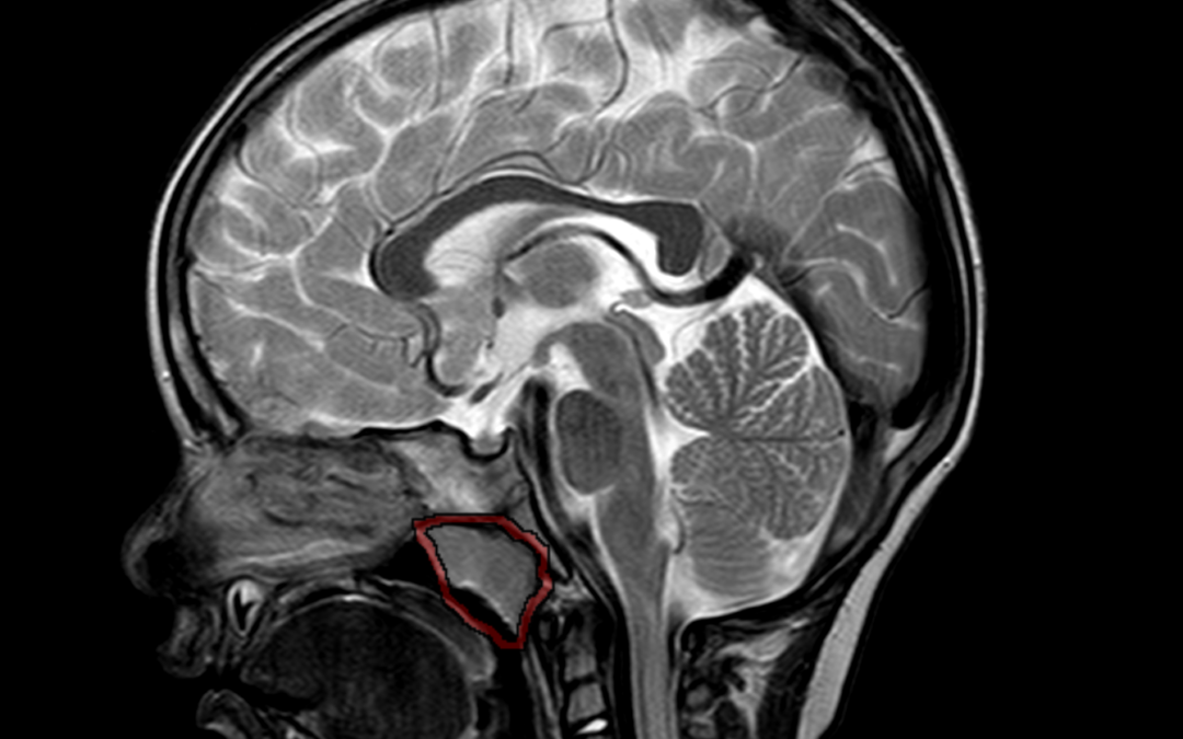
by Nithesh Ravindran | Mar 15, 2023
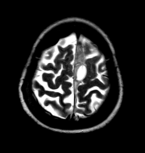
Case courtesy : Dr.Cicy Kuncherian,Welcare Hospital.
Age: 27 Yrs
Sex: Male
Complaints: Swelling frontal region.
Case study: A well defined soft tissue density lesion (~ +13HU) measuring ~13 x 11 mm noted in the frontal region involving subgaleal layer. No evidence of intracranial invasion / bone erosion.
Images:

Conclusion :A well defined soft tissue lesion involving the mid frontal region causing superficial skin asymmetry as described – Imaging features in favour of dermoid cyst. Possible DD: Sebaceous cyst.
by Nithesh Ravindran | Dec 4, 2022
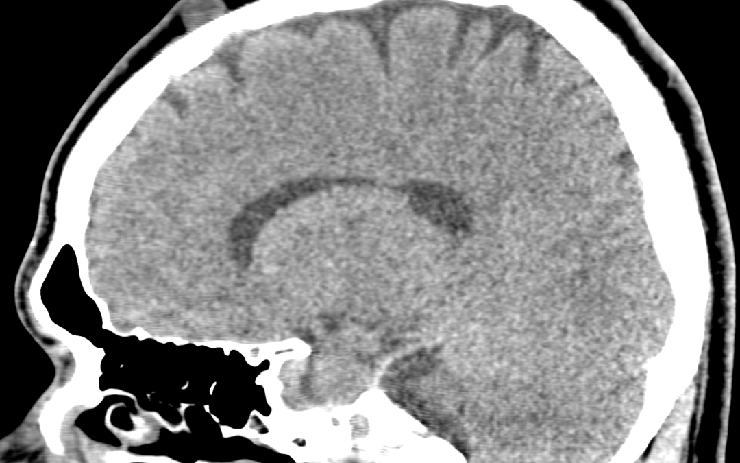
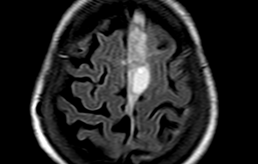
Case courtesy : Dr. Sreedevi Varun DMRD DNB, Sunrise Hospital
Age: 77Yrs
Sex: Female
Complaints: H/o fall.
Case study:
Area of heterogeneous T2 hyperintense signal in midline falx with maximum diameter 14 mm in highfrontal region. Cyst like area measuring 26 x 24 mm noted in midline along falx.Altered signal intensity of adjacent frontal lobe bilaterally – likely edema.
Thin extra axial hyperintense signal along left parieto-temporal convexity with maximum diameter 3 mm.
Images:
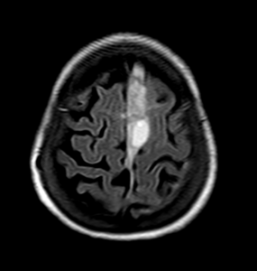
Conclusion :
Acute subdural hemorrhage in midline falx (14 mm in maximum diameter).
Thin subdural hemorrhage along left parieto-temporal convexity.
Hyperintense signal in bilateral frontal lobe adjacent to SDH – likely parenchymal edema.
by Nithesh Ravindran | Dec 4, 2022
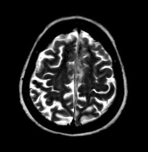
Case courtesy : Dr.Sreedevi Varun DMRD DNB, Sunrise Hospital
Age: 4Yrs
Sex: Male
Case study:
An extra axial CSF intensity lesion in left anterior temporal fossa measuring 3.3 x 1.3 x 3.5 cm (TR x AP x CC) with no significant mass effect or perilesional edema. Prominent bilateral retro cerebellar CSF intensity area measuring approximately 3.6 x 1.7 cm (TR x AP).
Images:
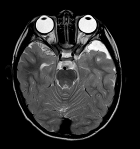
Conclusion :
Extra axial CSF intensity lesion in left anterior temporal fossa – favor arachnoid cyst.
Prominent cisterna magna.
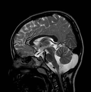
by Nithesh Ravindran | Dec 4, 2022
Age: 2Yrs
Sex: Male
Complaints: Speech Difficulty.
Case study:
Adenoid gland appears enlarged measuring ~ 16 .8mm (parasagittal). There is evident narrowing of nasopharyngeal air way.
Images:
Conclusion : Adenoid gland hypertrophy with nasopharyngeal airway narrowing.
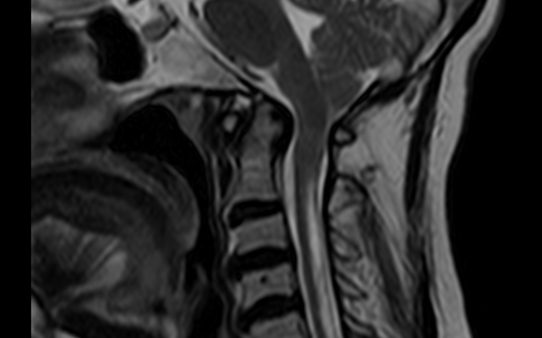
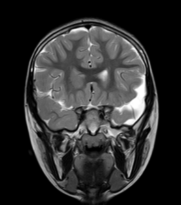
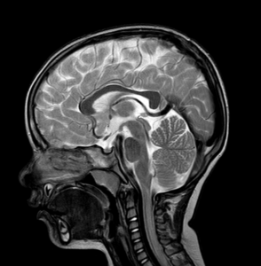
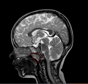
by Nithesh Ravindran | Dec 4, 2022
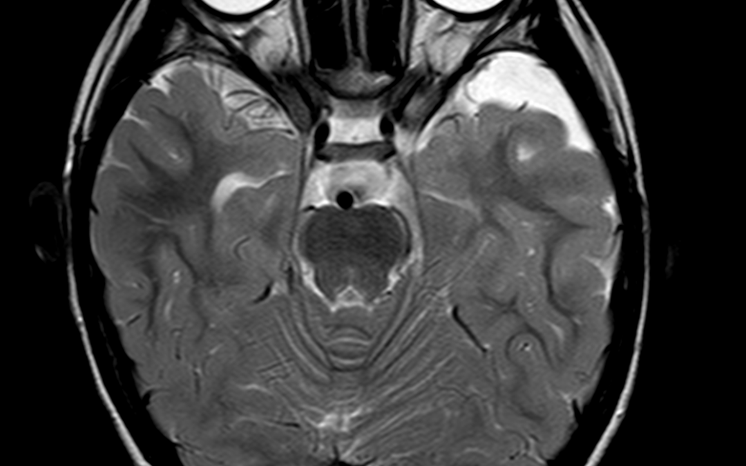
Case courtesy : Dr. Sameer Hyderali MD, Sunrise Hospital
Age: 55Yrs
Sex: Female
Case study:
Low placed cerebellar tonsils 5 mm below the foramen magnum.
Tight foramen magnum with effaced 4th ventricle and the cisternal spaces noted.
The cervico-medullary junction shows compression.
Hyperintense area seen within the cord posteriorly from C1 to C6 level.
Images:
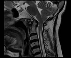
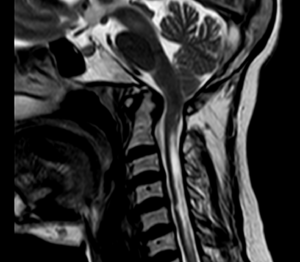
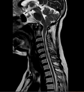
Conclusion :
Features consistent with Chiari 1 malformation with syringohydromyelia.
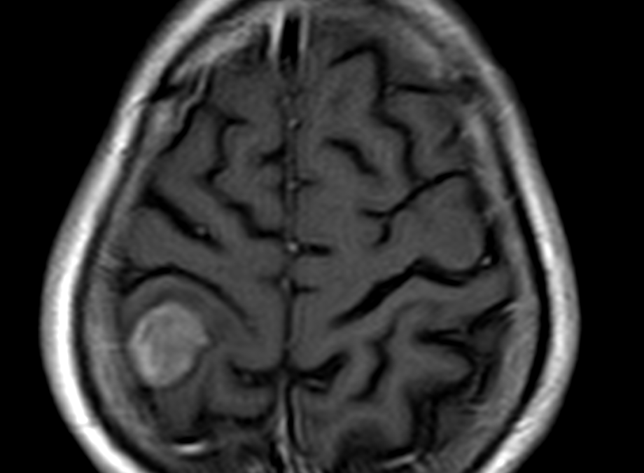
by Nithesh Ravindran | Oct 11, 2022
Age: 65Yrs
Sex: Male
Complaints: Head ache
Case study: Moderately enhancing right posterior parietal foci showing broad base to the dura measuring 1.8 x 3.4 x 2.5 cm. The foci shows evidence of dural tail with evidence of indentation on the adjacent brain parenchyma with mild adjacent white matter long TR hyperintensities, hypointense on T1WI. This foci shows mild diffusion restriction on the DWI/ADC.
Images:
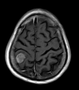
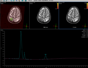
Conclusion : A right sided posterior parietal significantly enhancing foci, broad base to dura with dural tail and adjacent brain parenchymal indentation and mild parenchymal edema – To consider possibility of meningioma.
MR spectroscopy shows significant elevation of the choline peaks with significant reduction in the NAA and altered CHO/NAA ratio. Significantly reduced creatine ratios also noted.