by Nithesh Ravindran | Oct 11, 2022
Age: 45Yrs
Sex: Female
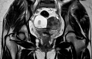
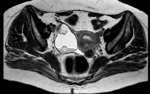
Case study: Uterus: Normal in size and shows two separate uteri with widely divergent apices, two separate cervices noted. Normal uterine zone anatomy is preserved in each uteri. Mildly heterogeneous myometrium noted in the right uteri with junctional zone measuring 10 mm. Intramural fibroid measuring 10 x 9 mm noted in the medial wall of left uteri.
Images:
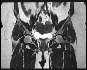
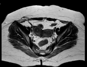
Conclusion : Imaging features are suggestive of uterine didelphys with intramural fibroid in left uteri and heterogeneous myometrium of right uteri (? Adenomyosis) as described.
by Nithesh Ravindran | Oct 11, 2022
Age: 41Yrs
Sex: Female
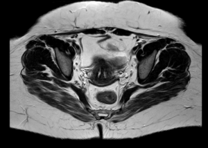
Case Study: Well-defined T2/STIR hyperintense signal cystic lesion in the postero-lateral aspect of vagina.
Images:
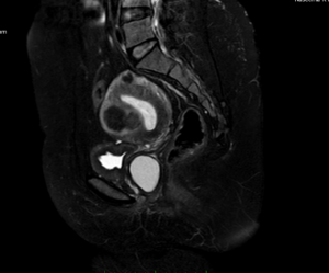
Conclusion : Postero-lateral wall of vagina shows a cyst as described – Suggestive of likely Bartholin’s cyst.
by Nithesh Ravindran | Aug 24, 2022
Age: 41 Yrs
Sex: Female
Complaints: Right lower abdominal pain.
Images:


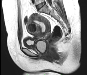
Case study: A large heterogenous exophytic lesion measuring 6.3 x 4.9 cm seen in the right adnexa derived from the ovary showing T1 & T2 hyperintense signal intensity and suppression in the STIR sequences. The lesion exhibits irregular solid nodular areas within. Other findings – leiomyomas.
Conclusion: Right adnexal lesion showing typical MRI features of T1/T2 hyperintensity and loss of signal in Fat suppressed sequences with nodular areas suggesting fat contents. – Dermoid cyst of the ovary. Variant – Matured cystic teratoma.
Reference : 1.https://radiopaedia.org/articles/mature-cystic-ovarian-teratoma-1 Mature cystic teratomas are encapsulated tumors with mature tissue or organ components. They are composed of well-differentiated derivations from at least two of the three germ cell layers (i.e. ectoderm, mesoderm, and endoderm). They, therefore, contain developmentally mature skin complete with hair follicles and sweat glands, sometimes luxuriant clumps of long hair, and often pockets of sebum, blood, fat (93%) 10, bone, nails, teeth, eyes, cartilage, and thyroid tissue. Typically their diameter is smaller than 10 cm, and rarely more than 15 cm. Real organoid structures (teeth, fragments of bone) may be present in ~30% of cases.