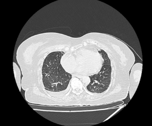
by Nithesh Ravindran | Mar 15, 2023
Case courtesy : Dr. Cicy Kuncheria Welcare Hospital
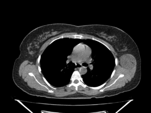
Age: 61Yrs
Sex: Female
Complaints: Recurrent pain over left mid-clavicular region.
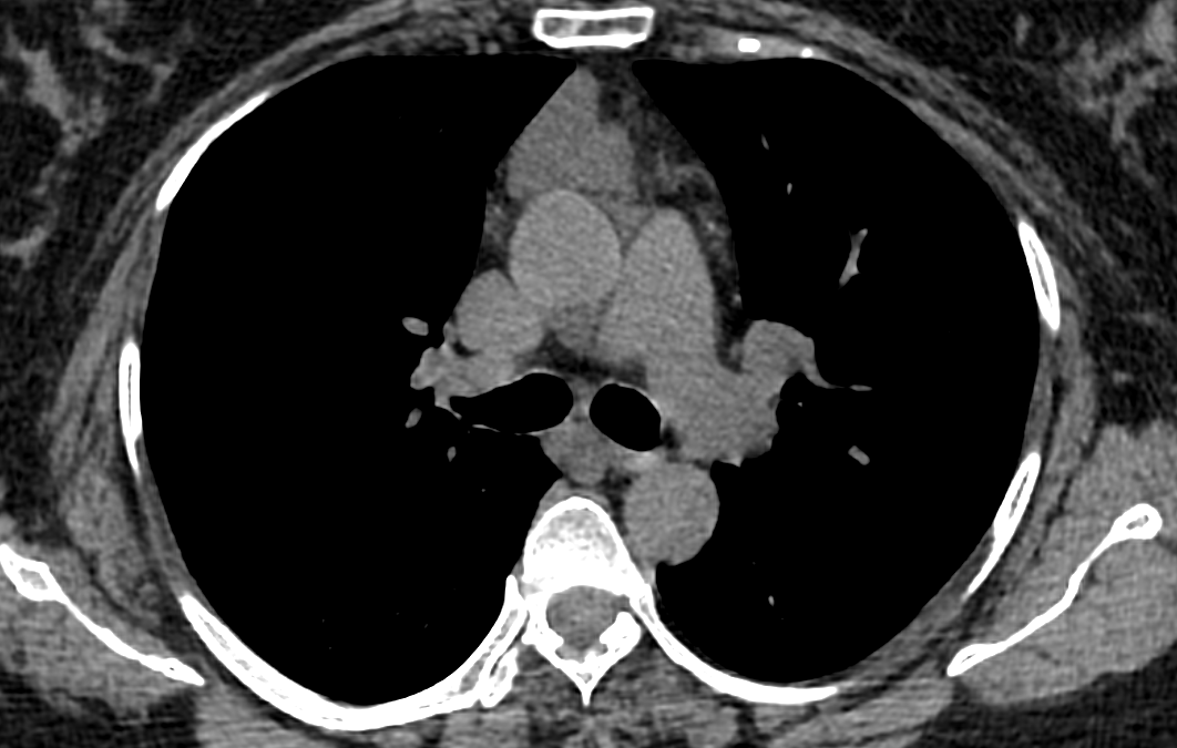
Case study: A smooth well defined soft tissue mass with fluid attenuation in the anterior mediastinum measuring ~3.9 x 2.9 x 3.4cm (TR XAP XCC).The lesion shows homogenous density.No calcification.
Images:
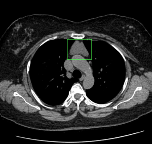
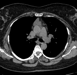
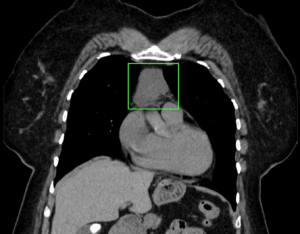
Conclusion : Smooth well defined anterior mediastinal mass lesion with fluid attenuation -imaging features are in favor of cystic mediastinal mass lesion. DD: Thymic cyst
by Nithesh Ravindran | Oct 14, 2022
Credits : Dr.Nithin
Age: 89 Yrs
Sex: Female
Complaints:
Case study: Cluster of cystic spaces of variable size with air fluid level noted in the upper lobe of right lung.
Right mild pleural effusion noted.
No evidence of any compensatory lung hyperinflation.
Images:
Cystic dilatation of bronchioles in the right upper lobe.
Right pleural effusion
Conclusion: Cystic bronchiectasis with air fluid level involving the right upper lobe.
Right pleural effusion.
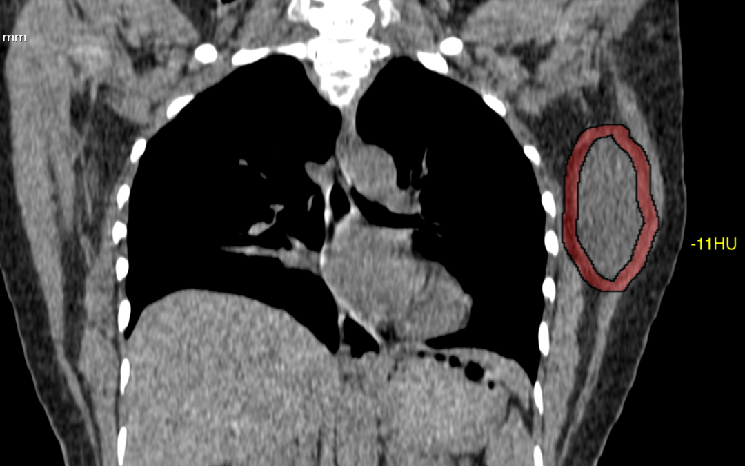
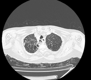
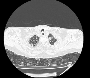
by Nithesh Ravindran | Dec 15, 2021
Age: 48 Yrs
Sex: Female
Complaints: H/o left chest wall swelling. No pain or skin abnormality.
Images:
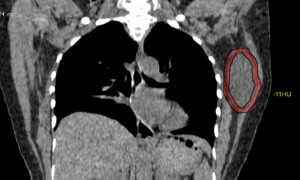
Well circumscribed hypo to isodense (max. -15HU) oval shaped mass lesion measuring ~6.6 x 4.0 x 3.8 cm (CC x TR x AP) noted evolving from the left latissimus dorsi and muscle involving the intramuscular space between subscapularis. No evidence of any bone involvement.
Case Discussion :
Well defined smooth solid hypo to iso-dense lesion in the left chest wall seen arising from the latissimus dorsi muscle.
The is lesion is seen as a solid mass with hypo-dense attenuation, however the lesion shows typical fatty appearance.
The report was given as chest wall mass arising from left latissimus dorsi muscle.
Possible DD’s include: 1. Lipoma. 2. Liposarcoma 3.Schwannoma.
Conclusion: FNA done. Histopathology came as fat containing lesion and suggested Excision biopsy.
Reference
- Lee JH, Do HD, Lee JC. Well-circumscribed type of intramuscular lipoma in the chest wall. Journal of cardiothoracic surgery. 8: 181. doi:10.1186/1749-8090-8-181 – Pubmed
- 3. López Soriano A, Tomasello A, Luburich P, Noel A. Fat necrosis in a chest wall lipoma. AJR. American journal of roentgenology. 183 (3): 866. doi:10.2214/ajr.183.3.1830866 – Pubmed
by Nithesh Ravindran | Dec 14, 2021
Name: Gowrikutty
Age: 80 Yrs
Sex: Female
Complaints:
Introduction:
Case study: Diffuse degenerative changes in the form of flowing osteophytes in the anterior aspect of thoracic vertebrae with loss of disc space and fusion of multiple vertebral bodies.
Images
Conclusion: Diffuse degenerative changes in the thoracic vertebrae in the form of flowing osteophytes, disc space reduction and fusion of multiple vertebrae bodies. – Features in favour of diffuse idiopathic skeletal hyperostosis (DISH).
Reference :