by Nithesh Ravindran | Dec 4, 2022
Age: 2Yrs
Sex: Male
Complaints: Speech Difficulty.
Case study:
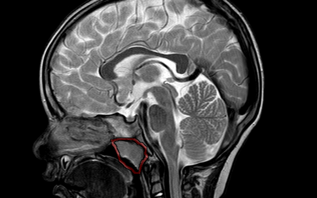
Adenoid gland appears enlarged measuring ~ 16 .8mm (parasagittal). There is evident narrowing of nasopharyngeal air way.
Images:
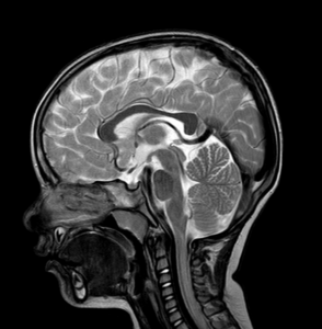
Conclusion : Adenoid gland hypertrophy with nasopharyngeal airway narrowing.
by Nithesh Ravindran | Dec 4, 2022
Case courtesy : Dr Sajith Selvaganesan MD FSCCT FSCMR, Sunrise Hospital
Age: 68 Yrs
Sex: Female
Case study:
High grade partial tear of subscapularis tendon with no retraction of muscle.
Altered intra-substance signal intensity noted in the supraspinatus tendon reaching up to the articular surface.
Altered signal intensity noted in the posterolateral aspect of humeral head.
Type V superior labral anterior posterior tear noted with minimal fluid along the interface.
Type II acromion seen.
Minimal joint effusion with subcoracoid bursal fluid.
Images:
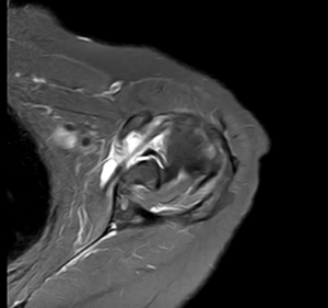
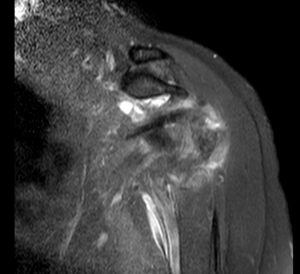
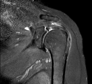
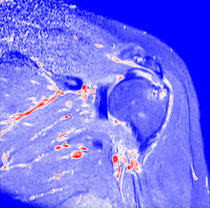
Conclusion :
High grade partial tear of subscapularis tendon.
Partial tear of supraspinatus tendon.
Type V superior labral anterior posterior tear.
Altered signal intensity in the posterolateral aspect of humeral head – Likely hillsach’s lesion.
Minimal joint effusion with subcoracoid bursal fluid.
Acromioclavicular joint athropathy changes.
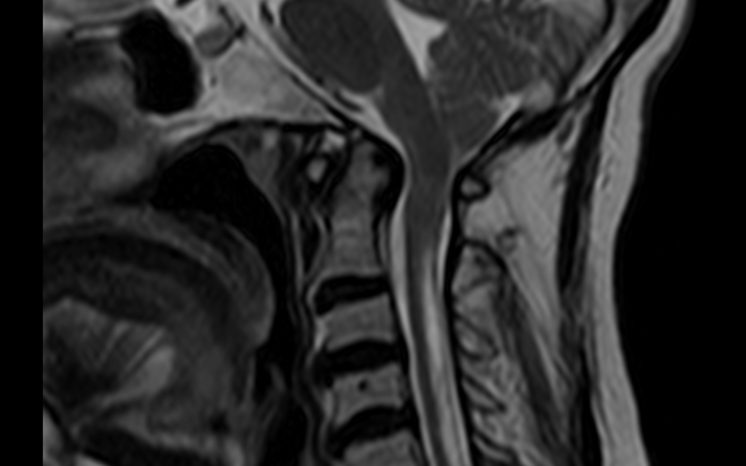
by Nithesh Ravindran | Dec 4, 2022
Case courtesy : Dr. Sameer Hyderali MD, Sunrise Hospital
Age: 55Yrs
Sex: Female
Case study:
Low placed cerebellar tonsils 5 mm below the foramen magnum.
Tight foramen magnum with effaced 4th ventricle and the cisternal spaces noted.
The cervico-medullary junction shows compression.
Hyperintense area seen within the cord posteriorly from C1 to C6 level.
Images:
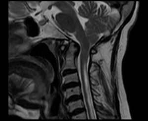
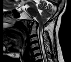
Conclusion :
Features consistent with Chiari 1 malformation with syringohydromyelia.
by Nithesh Ravindran | Oct 14, 2022
Credits : Dr.Nithin
Age: 89 Yrs
Sex: Female
Complaints:
Case study: Cluster of cystic spaces of variable size with air fluid level noted in the upper lobe of right lung.
Right mild pleural effusion noted.
No evidence of any compensatory lung hyperinflation.
Images:
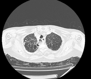
Cystic dilatation of bronchioles in the right upper lobe.
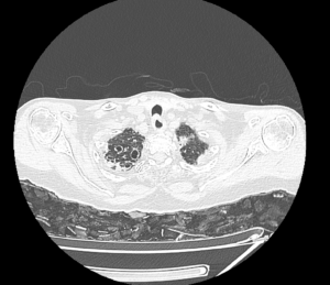
Right pleural effusion
Conclusion: Cystic bronchiectasis with air fluid level involving the right upper lobe.
Right pleural effusion.
by Nithesh Ravindran | Oct 11, 2022
Age: 45Yrs
Sex: Female
Case study: Uterus: Normal in size and shows two separate uteri with widely divergent apices, two separate cervices noted. Normal uterine zone anatomy is preserved in each uteri. Mildly heterogeneous myometrium noted in the right uteri with junctional zone measuring 10 mm. Intramural fibroid measuring 10 x 9 mm noted in the medial wall of left uteri.
Images:
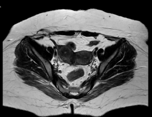
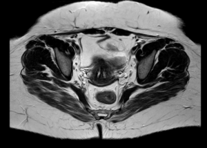
Conclusion : Imaging features are suggestive of uterine didelphys with intramural fibroid in left uteri and heterogeneous myometrium of right uteri (? Adenomyosis) as described.
by Nithesh Ravindran | Oct 11, 2022
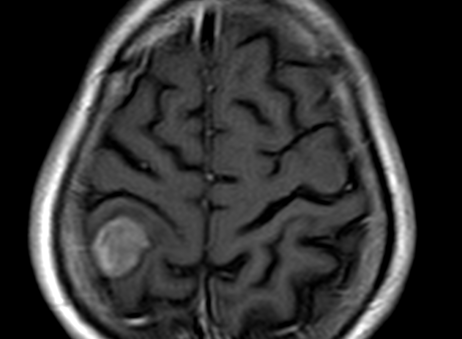
Age: 65Yrs
Sex: Male
Complaints: Head ache
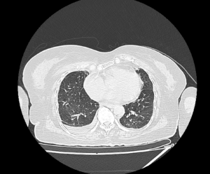
Case study: Moderately enhancing right posterior parietal foci showing broad base to the dura measuring 1.8 x 3.4 x 2.5 cm. The foci shows evidence of dural tail with evidence of indentation on the adjacent brain parenchyma with mild adjacent white matter long TR hyperintensities, hypointense on T1WI. This foci shows mild diffusion restriction on the DWI/ADC.
Images:
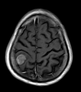
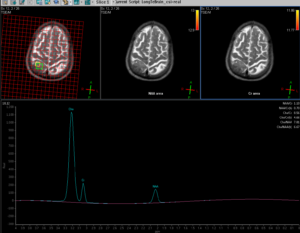
Conclusion : A right sided posterior parietal significantly enhancing foci, broad base to dura with dural tail and adjacent brain parenchymal indentation and mild parenchymal edema – To consider possibility of meningioma.
MR spectroscopy shows significant elevation of the choline peaks with significant reduction in the NAA and altered CHO/NAA ratio. Significantly reduced creatine ratios also noted.