by Nithesh Ravindran | Dec 18, 2021
Name: Sasidharan
Age: 67 Yrs
Sex: Male
Complaints: To r/o right inguinal hernia
Introduction:
Case study: An abdominal wall defect measuring ~25 mm noted in the right inguinal region with herniation of omental fat and bowel loops as content. Cough impulse positive.
The herniated bowel loop seen extending in to the sub inguinal region, however no bowel contents in the ipsilateral hemiscrotum.
Images
Conclusion: Right inguinal hernia.
by Nithesh Ravindran | Dec 18, 2021
Name: Guljar Hussain
Age: 28 Yrs
Sex: Male
Complaints: H/o lower abdominal pain.
Introduction:
Case study: An abdominal wall defect measuring ~33 mm noted in the right inguinal region with herniation of omental fat and bowel loop as content extending into the inguinal canal and reaching into the ipsilateral scrotal sac.
Images
Conclusion: Right inguino-scrotal hernia
by Nithesh Ravindran | Dec 16, 2021
Age: 48 Yrs
Sex: Female
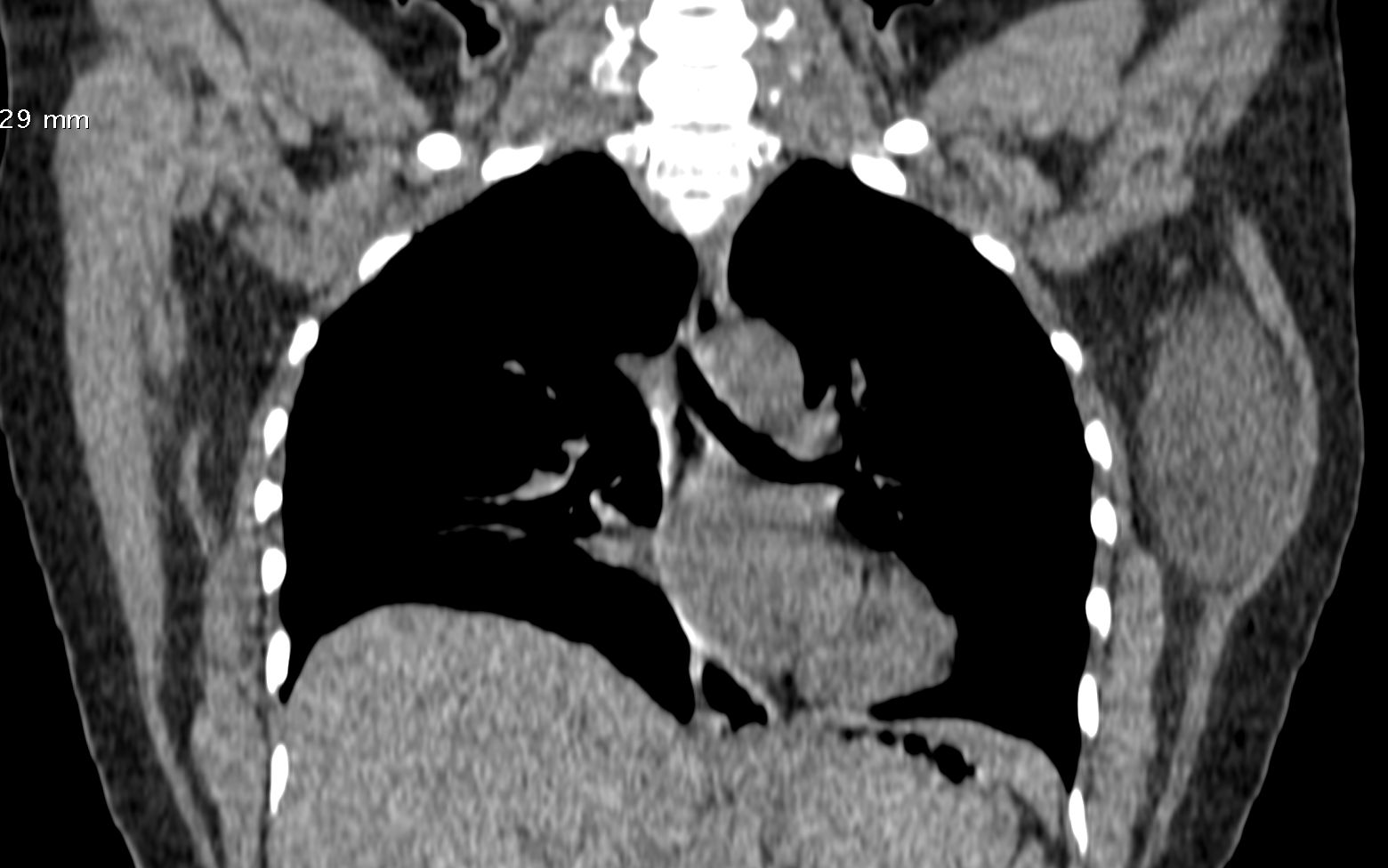
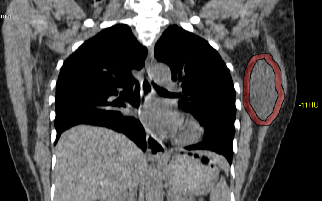
Complaints: H/o left chest wall swelling. No pain or skin abnormality.
Screen Shot 2021-12-15 at 19.37.26
Screen Shot 2021-12-15 at 19.38.33
Case Discussion :
Well defined smooth solid hypo to isodense lesion in the left chest wall seen arising from the latissimus dorsi muscle.
The is lesion is seen as a solid mass with hypo-dense attenuation, however the lesion shows typical fatty appearance.
The report was given as chest wall mass arising from left latissimus dorsi muscle.
Possible DD’s include: 1. Lipoma. 2. Schwannoma.
by Nithesh Ravindran | Dec 15, 2021
Name: Podiyamma
Age: 72 Yrs Sex: Female
Complaints: To r/o renal calculi. Introduction:
Images
Case study: RIF: Mild probe tenderness present. Appendix visualized enlarged, measures ~10 mm and shows a calculus measuring ~5.3 mm at the tip. Periappendiceal inflammatory changes noted in the form of wall edema and minimal fluid. Images Conclusion: Appendix appears enlarged with surrounding inflammatory changes and a calculus at the tip. – Features in favour of acute appendicitis with appendicolith. Suggested BRE and CRP correlation.
Reference :
by Nithesh Ravindran | Dec 15, 2021
Age: 48 Yrs
Sex: Female
Complaints: H/o left chest wall swelling. No pain or skin abnormality.
Images:
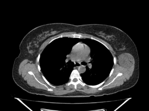
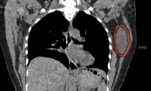
Well circumscribed hypo to isodense (max. -15HU) oval shaped mass lesion measuring ~6.6 x 4.0 x 3.8 cm (CC x TR x AP) noted evolving from the left latissimus dorsi and muscle involving the intramuscular space between subscapularis. No evidence of any bone involvement.
Case Discussion :
Well defined smooth solid hypo to iso-dense lesion in the left chest wall seen arising from the latissimus dorsi muscle.
The is lesion is seen as a solid mass with hypo-dense attenuation, however the lesion shows typical fatty appearance.
The report was given as chest wall mass arising from left latissimus dorsi muscle.
Possible DD’s include: 1. Lipoma. 2. Liposarcoma 3.Schwannoma.
Conclusion: FNA done. Histopathology came as fat containing lesion and suggested Excision biopsy.
Reference
- Lee JH, Do HD, Lee JC. Well-circumscribed type of intramuscular lipoma in the chest wall. Journal of cardiothoracic surgery. 8: 181. doi:10.1186/1749-8090-8-181 – Pubmed
- 3. López Soriano A, Tomasello A, Luburich P, Noel A. Fat necrosis in a chest wall lipoma. AJR. American journal of roentgenology. 183 (3): 866. doi:10.2214/ajr.183.3.1830866 – Pubmed