by Nithesh Ravindran | Aug 28, 2022
Age: 27yr
Sex: Female
Complaints: Gait ataxia and ophthalmoplegia confusion.
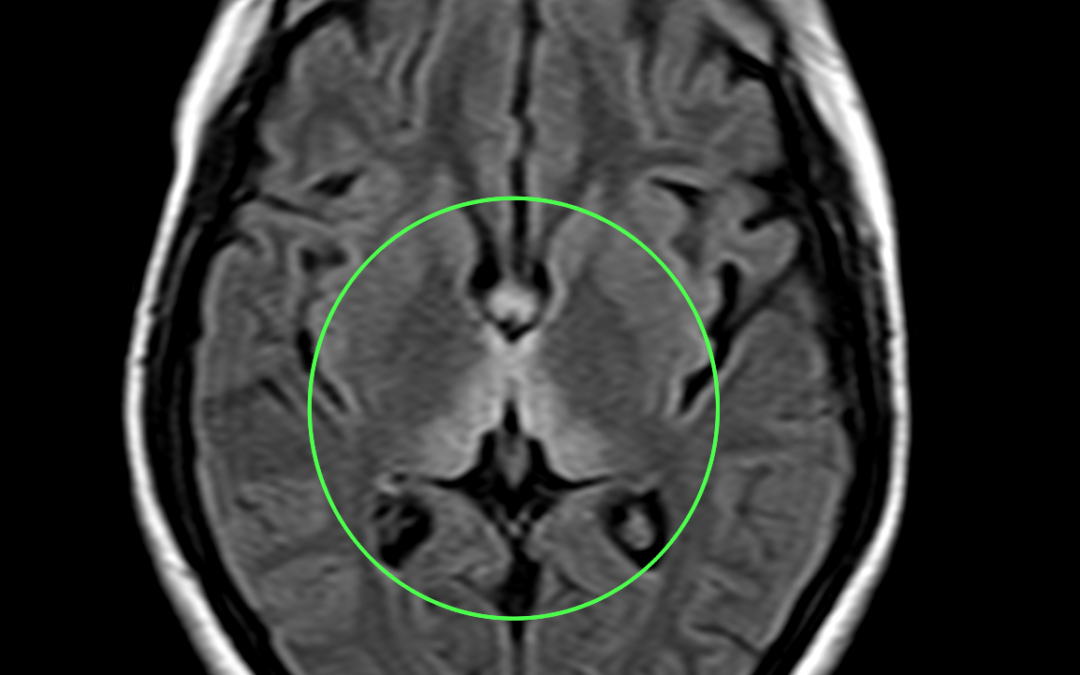
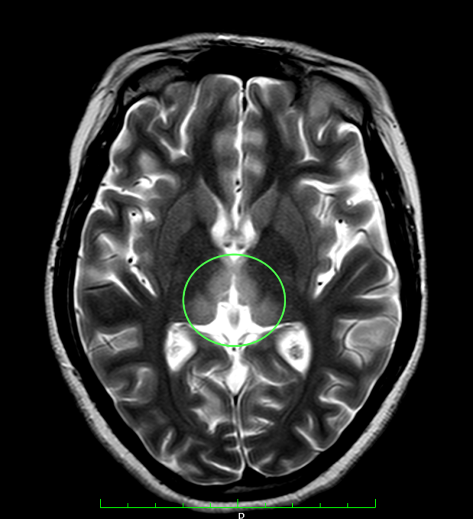
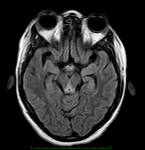
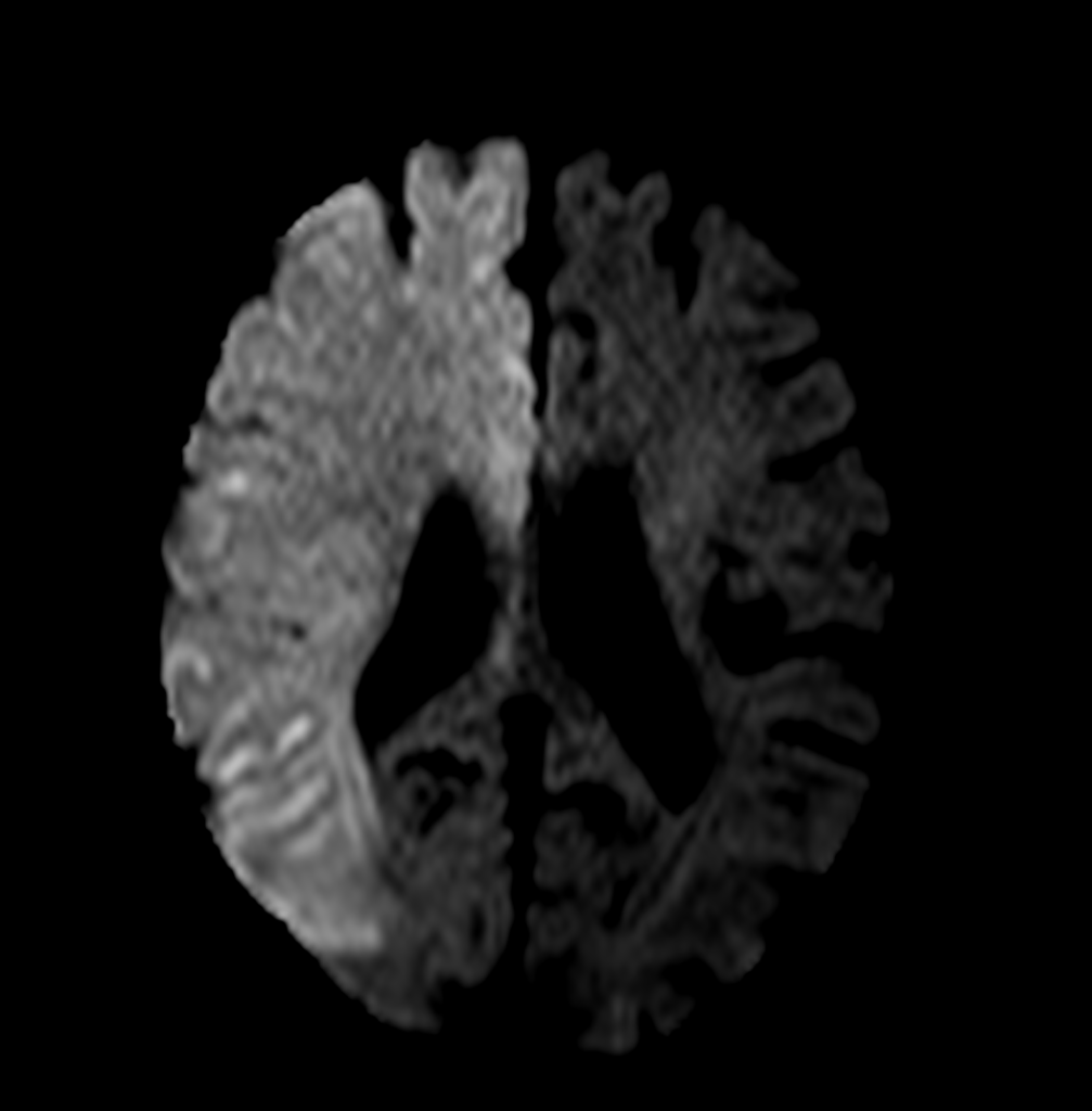
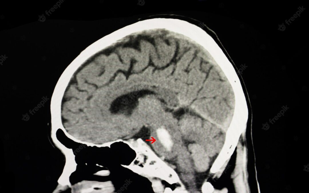
Case study: Areas of symmetrical increased T2/FLAIR signal seen involving dorsomedial thalami, tectal plate, periaqueductal area, around 3rd ventricle, mammillary bodies, posterior medulla (in medial vestibular nuclei and hypoglossal nuclei) with corresponding subtle diffusion restriction.
Images:
T2WI shows hyperintense signal involving dorsomedial aspect of thalamus.

FLAIR sequence shows hyperintensity involving the medial aspect of thalamus
Conclusion : Imaging features suggestive of wernicke encephalopathy.
by Nithesh Ravindran | Aug 28, 2022
Age: 74yr
Sex: Female
Complaints: Left limb weakness.
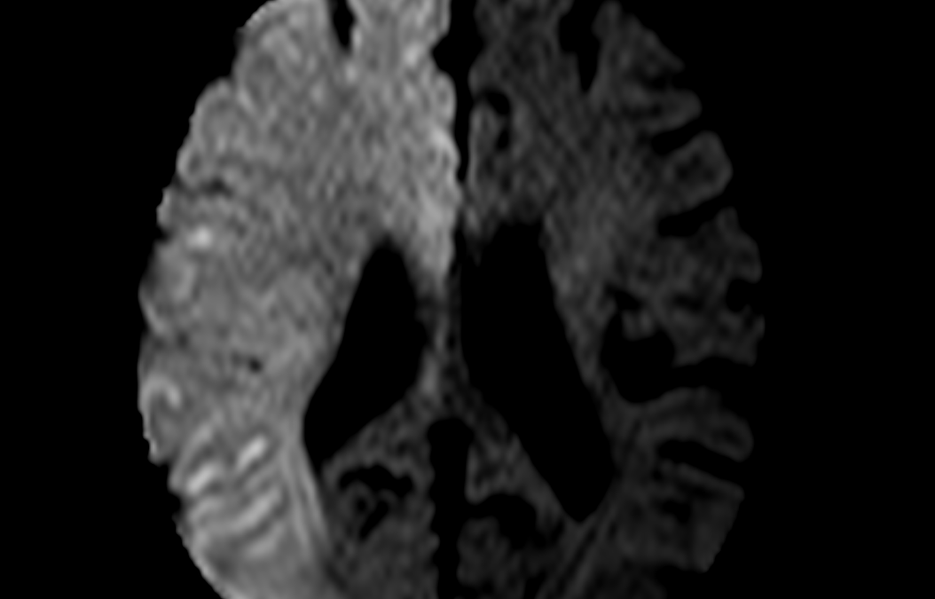
Case study: Large area of diffusion restriction with corresponding low ADC signal seen involving right fronto- parieto- temporal lobe with no signal abnormalities in corresponding T2 and FLAIR sequence.Angiography study shows non-visualisation right internal carotid artery (ICA) and its branches.No evidence of blooming in GRE sequence.
Images:
DWI
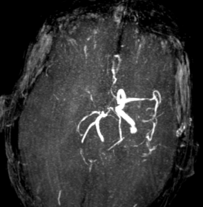
Angiography

ADC
Conclusion :
- Acute infract involving right MCA and ACA territory.
- Mild atrophic changes in brain.
- MR angiogram (cerebral angiogram) shows non visualized right ICA and its main branches indicating significant thrombosis.
.
by Nithesh Ravindran | Aug 24, 2022
Age: 41 Yrs
Sex: Female
Complaints: Right lower abdominal pain.
Images:

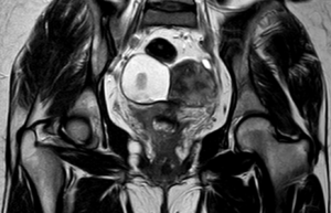
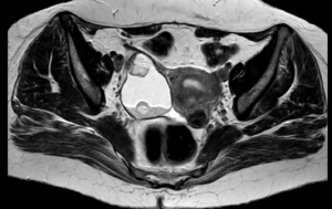

Case study: A large heterogenous exophytic lesion measuring 6.3 x 4.9 cm seen in the right adnexa derived from the ovary showing T1 & T2 hyperintense signal intensity and suppression in the STIR sequences. The lesion exhibits irregular solid nodular areas within. Other findings – leiomyomas.
Conclusion: Right adnexal lesion showing typical MRI features of T1/T2 hyperintensity and loss of signal in Fat suppressed sequences with nodular areas suggesting fat contents. – Dermoid cyst of the ovary. Variant – Matured cystic teratoma.
Reference : 1.https://radiopaedia.org/articles/mature-cystic-ovarian-teratoma-1 Mature cystic teratomas are encapsulated tumors with mature tissue or organ components. They are composed of well-differentiated derivations from at least two of the three germ cell layers (i.e. ectoderm, mesoderm, and endoderm). They, therefore, contain developmentally mature skin complete with hair follicles and sweat glands, sometimes luxuriant clumps of long hair, and often pockets of sebum, blood, fat (93%) 10, bone, nails, teeth, eyes, cartilage, and thyroid tissue. Typically their diameter is smaller than 10 cm, and rarely more than 15 cm. Real organoid structures (teeth, fragments of bone) may be present in ~30% of cases.
by Nithesh Ravindran | Jan 27, 2022
Age: 68 Yrs
Sex: Female
Complaints: Spontaneous headache.
Case study: A well defined hyperdense (max. ~60HU) area measuring ~19 x 16 x 6 mm (TR x CC x AP) noted in the left pontine hemisphere extending – Features in favor of acute pontine hemorrhage. No evidence of mass effect/hydrocephalus/intra-ventricular hemorrhage.
Conclusion: Acute pontine hemorrhage . – Features in favor of hypertensive etiology.
by Nithesh Ravindran | Jan 27, 2022
Name: Aneesh
Age: 28 Yrs
Sex: Male
Complaints:
Introduction:
Case study: A well-defined irregular non-enhancing hypodense cystic lesion measuring ~ 6.0 x 5.7 x 5.0 cm (CC x TR x AP) showing fluid attenuation (ranging +40 to -22HU) noted closely adjacent to the lower pole of right kidney and extending along the course of proximal ureter caudally for ~ 6.7 cm and significantly displacing it posteriorly.
The lesion is seen abutting the adjacent bowel loop superiorly and closely near to the psoas muscle posteriorly.
Images
Conclusion: Well-defined irregular non-enhancing hypodense cystic lesion in the right perirenal space and extending along the proximal ureter as described. – Features favor’s the possibility of Urinoma in the background of obstructive renal pathology. Suggested Aspiration cytology.
Reference :