by Nithesh Ravindran | Oct 14, 2022
Credits : Dr.Nithin
Age: 89 Yrs
Sex: Female
Complaints:
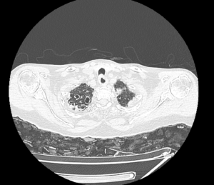
Case study: Cluster of cystic spaces of variable size with air fluid level noted in the upper lobe of right lung.
Right mild pleural effusion noted.
No evidence of any compensatory lung hyperinflation.
Images:
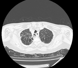
Cystic dilatation of bronchioles in the right upper lobe.
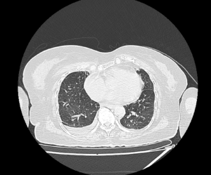
Right pleural effusion
Conclusion: Cystic bronchiectasis with air fluid level involving the right upper lobe.
Right pleural effusion.
by Nithesh Ravindran | Oct 11, 2022
Age: 45Yrs
Sex: Female
Case study: Uterus: Normal in size and shows two separate uteri with widely divergent apices, two separate cervices noted. Normal uterine zone anatomy is preserved in each uteri. Mildly heterogeneous myometrium noted in the right uteri with junctional zone measuring 10 mm. Intramural fibroid measuring 10 x 9 mm noted in the medial wall of left uteri.
Images:
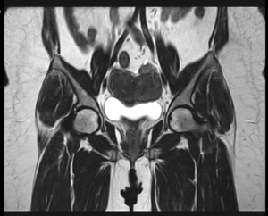
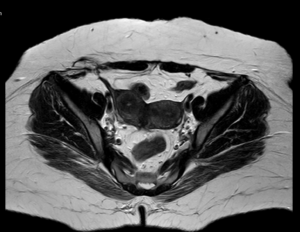
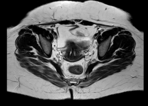
Conclusion : Imaging features are suggestive of uterine didelphys with intramural fibroid in left uteri and heterogeneous myometrium of right uteri (? Adenomyosis) as described.
by Nithesh Ravindran | Oct 11, 2022
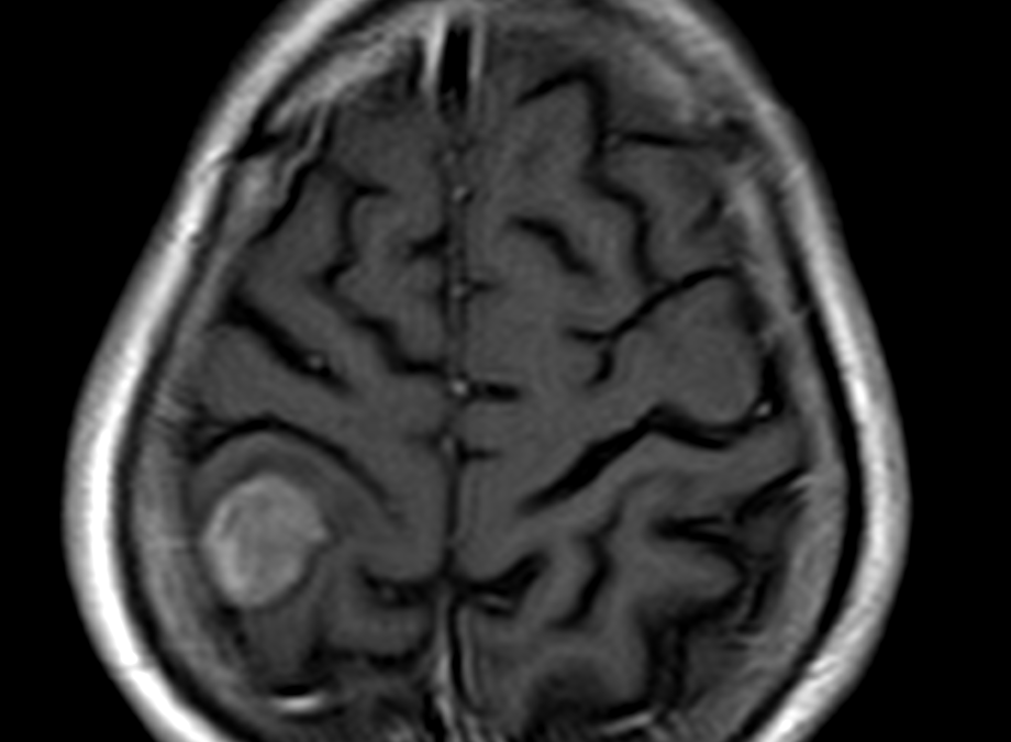
Age: 65Yrs
Sex: Male
Complaints: Head ache
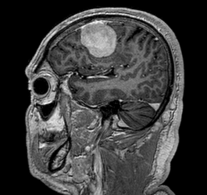
Case study: Moderately enhancing right posterior parietal foci showing broad base to the dura measuring 1.8 x 3.4 x 2.5 cm. The foci shows evidence of dural tail with evidence of indentation on the adjacent brain parenchyma with mild adjacent white matter long TR hyperintensities, hypointense on T1WI. This foci shows mild diffusion restriction on the DWI/ADC.
Images:
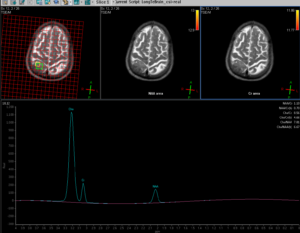
Conclusion : A right sided posterior parietal significantly enhancing foci, broad base to dura with dural tail and adjacent brain parenchymal indentation and mild parenchymal edema – To consider possibility of meningioma.
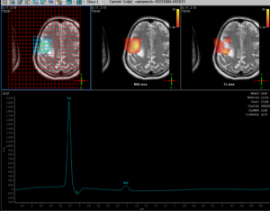
MR spectroscopy shows significant elevation of the choline peaks with significant reduction in the NAA and altered CHO/NAA ratio. Significantly reduced creatine ratios also noted.
by Nithesh Ravindran | Oct 11, 2022
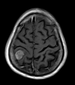
Age: 59yrs
Sex: Male
Complaints:
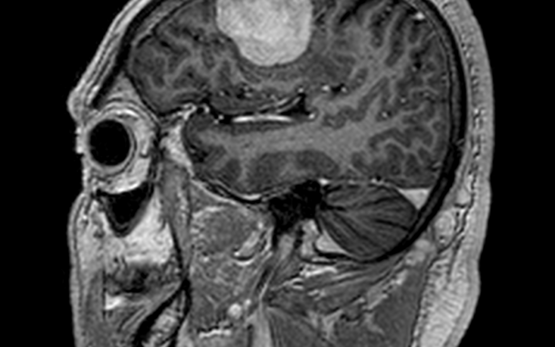
Case study: Supratentorial extra-axial dural based well defined lesion in right frontal convexity measuring 4.2 x 2.6 x 3.5 cm (AP x TR x CC) causing moderate perilesional edema and mild mass effect on adjacent brain parenchyma seen.
Dural tail and homogeneous vivid enhancement seen following contrast. The lesion appears iso intense to grey matter in T1WI and hyperintense in FLAIR/T2.
Images:
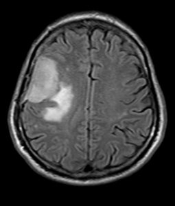
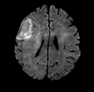
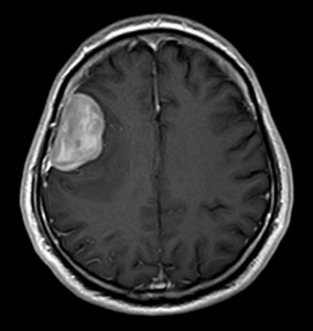
Conclusion :Extra-axial dural based vividly enhancing lesion in right frontal convexity causing moderate perilesional edema and mild mass effect – Imaging features favor meningioma.Mild diffusion restriction seen (hyperintense in DWI with intermediate signal in ADC). No blooming to suggest hemorrhage or calcification. MRS shows choline peak.
by Nithesh Ravindran | Oct 11, 2022
Age: 30Yrs
Sex: Male
Complaints: Pain on foot while walking.
Case study: Subchondral high signal noted in the mid aspect of the talus and the calcaneum with small subchondral cystic areas – Apparent on the STIR sequences. Evidence of osteophytic changes with cortical irregularity and narrowing of the talo-calcaneal joint anteriorly. The osteophyte is seen encroaching into the sinus tarsi with narrowing.
The tibio-calcaneal angle (kites angle) is 83 degrees – Normal.
The talo-calcaneal angle is 45 degrees – Abnormal.
Images:
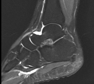
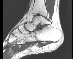
Conclusion : Subchondral talo-calcaneal high marrow signals with anterior joint space narrowing, osteophytic formation and encroachment into the sinus tarsi – Suggestive of talo-calcaneal impingement.
Increased talo-calcaneal angle (45 degrees) – Hindfoot valgus.
Peroneus longus and brevis tenosynovitis.