by Nithesh Ravindran | Dec 4, 2022
Case courtesy : Dr. Sreedevi Varun DMRD DNB, Sunrise Hospital
Age: 77Yrs
Sex: Female
Complaints: H/o fall.
Case study:
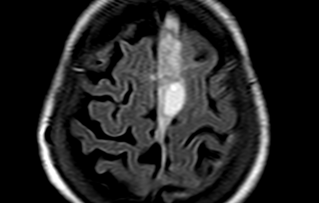
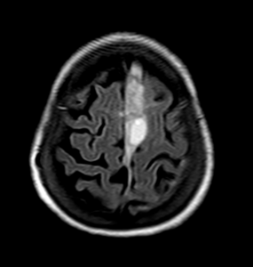
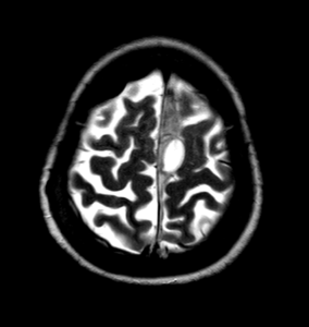
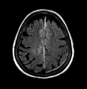
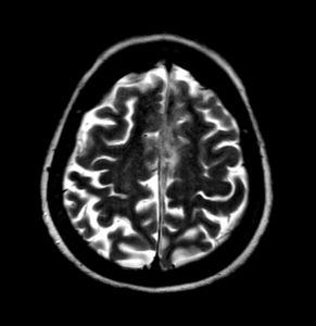
Area of heterogeneous T2 hyperintense signal in midline falx with maximum diameter 14 mm in highfrontal region. Cyst like area measuring 26 x 24 mm noted in midline along falx.Altered signal intensity of adjacent frontal lobe bilaterally – likely edema.
Thin extra axial hyperintense signal along left parieto-temporal convexity with maximum diameter 3 mm.
Images:
Conclusion :
Acute subdural hemorrhage in midline falx (14 mm in maximum diameter).
Thin subdural hemorrhage along left parieto-temporal convexity.
Hyperintense signal in bilateral frontal lobe adjacent to SDH – likely parenchymal edema.
by Nithesh Ravindran | Dec 4, 2022
Case courtesy : Dr.Sreedevi Varun DMRD DNB, Sunrise Hospital
Age: 4Yrs
Sex: Male
Case study:
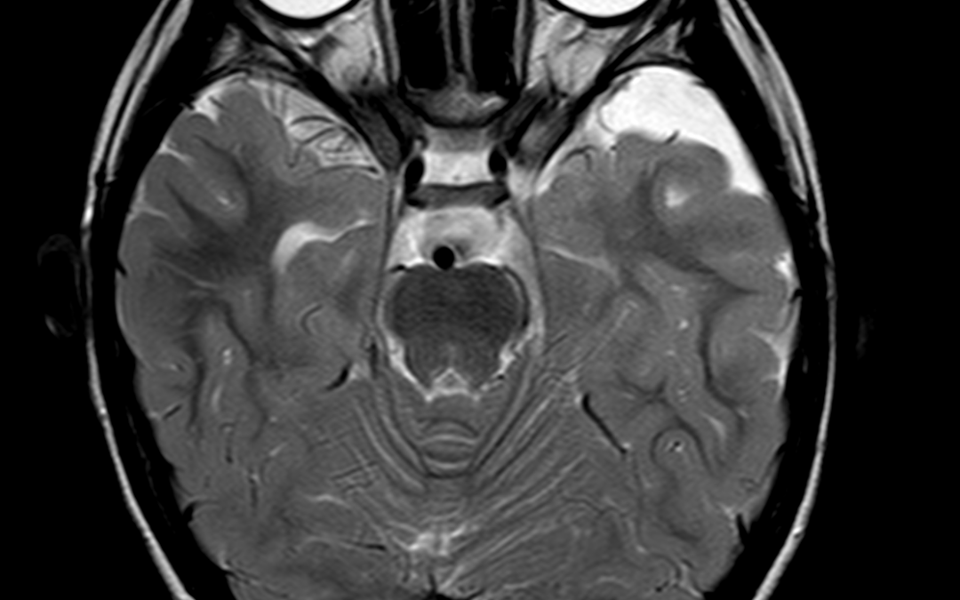
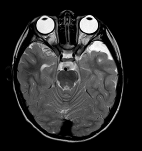
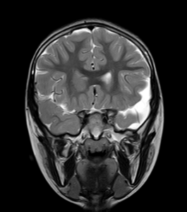
An extra axial CSF intensity lesion in left anterior temporal fossa measuring 3.3 x 1.3 x 3.5 cm (TR x AP x CC) with no significant mass effect or perilesional edema. Prominent bilateral retro cerebellar CSF intensity area measuring approximately 3.6 x 1.7 cm (TR x AP).
Images:
Conclusion :
Extra axial CSF intensity lesion in left anterior temporal fossa – favor arachnoid cyst.
Prominent cisterna magna.
by Nithesh Ravindran | Dec 4, 2022
Age: 2Yrs
Sex: Male
Complaints: Speech Difficulty.
Case study:
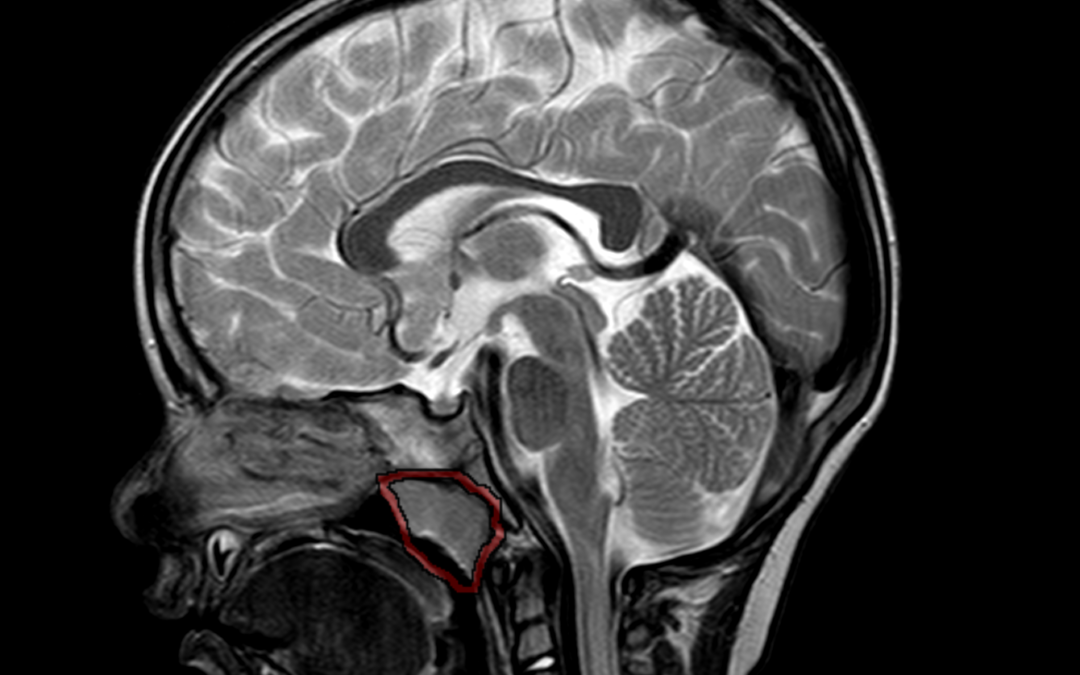
Adenoid gland appears enlarged measuring ~ 16 .8mm (parasagittal). There is evident narrowing of nasopharyngeal air way.
Images:
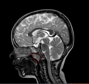
Conclusion : Adenoid gland hypertrophy with nasopharyngeal airway narrowing.
by Nithesh Ravindran | Dec 4, 2022
Case courtesy : Dr Sajith Selvaganesan MD FSCCT FSCMR, Sunrise Hospital
Age: 68 Yrs
Sex: Female
Case study:
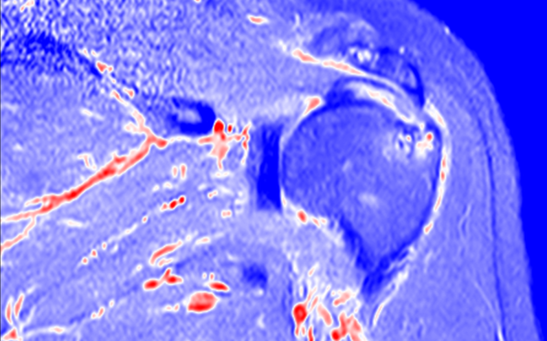
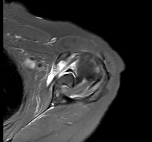
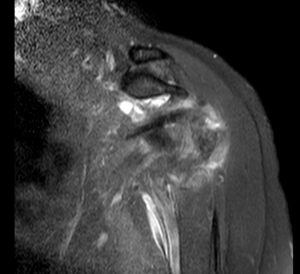
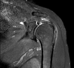
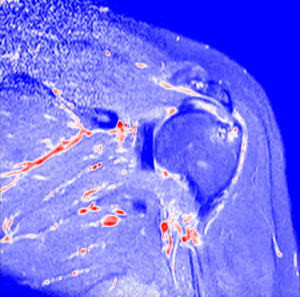
High grade partial tear of subscapularis tendon with no retraction of muscle.
Altered intra-substance signal intensity noted in the supraspinatus tendon reaching up to the articular surface.
Altered signal intensity noted in the posterolateral aspect of humeral head.
Type V superior labral anterior posterior tear noted with minimal fluid along the interface.
Type II acromion seen.
Minimal joint effusion with subcoracoid bursal fluid.
Images:
Conclusion :
High grade partial tear of subscapularis tendon.
Partial tear of supraspinatus tendon.
Type V superior labral anterior posterior tear.
Altered signal intensity in the posterolateral aspect of humeral head – Likely hillsach’s lesion.
Minimal joint effusion with subcoracoid bursal fluid.
Acromioclavicular joint athropathy changes.
by Nithesh Ravindran | Dec 4, 2022
Case courtesy : Dr. Sameer Hyderali MD, Sunrise Hospital
Age: 55Yrs
Sex: Female
Case study:
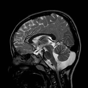
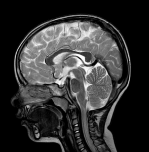
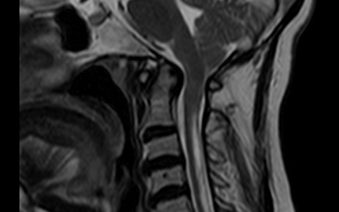
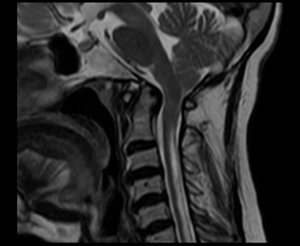
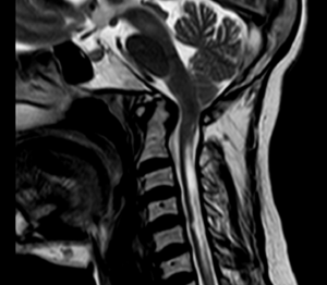
Low placed cerebellar tonsils 5 mm below the foramen magnum.
Tight foramen magnum with effaced 4th ventricle and the cisternal spaces noted.
The cervico-medullary junction shows compression.
Hyperintense area seen within the cord posteriorly from C1 to C6 level.
Images:
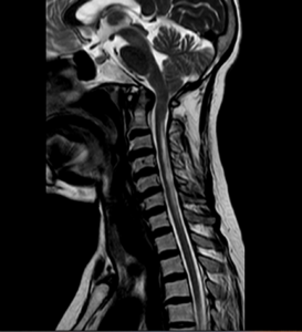
Conclusion :
Features consistent with Chiari 1 malformation with syringohydromyelia.