by Nithesh Ravindran | Dec 8, 2021
Mesenteric lymphadenopathy
Age: 3YR
Sex: Male
Complaints: H/o abdominal pain.
Introduction:
Case study:
Multiple enlarged mesenteric lymphnodes noted in the right iliac region, largest measuring ~15 x 6 mm with poorly maintained fatty hilum.
Images
Conclusion: Multiple enlarged right iliac lymphnodes. – Suggestive of lymphadenopathy.
Reference
by Nithesh Ravindran | Dec 7, 2021
Acute/subacute appendicitis
Age: 11
Sex: Male
Introduction:
Case study: RIF: Mild probe tenderness present. Appendix visualized measures ~8 mm with partially compressible lumen. Minimal free fluid collection noted adjacent to the tip of appendix measuring ~16 x 8 mm. No evidence of lymphadenopathy.
Images
Conclusion: Appendix appears enlarged with minimal surrounding fluid collection and mild probe tenderness. – Features suggestive of acute/subacute appendicitis. Suggested BRE correlation
Reference

by Nithesh Ravindran | Oct 27, 2021
Transitional cell carcinoma (TCC), also called urothelial cell carcinoma (UCC) of the bladder, is the most common primary neoplasm of the urinary bladder, and bladder TCC is the most common tumour of the entire urinary system.
CASE DISCUSSION
The bladder is by far the most common site of transitional cell carcinomas, 50 times more common than TCC of the renal pelvis, and 100 times more common than TCC of the ureter 1. Bladder TCCs are the most common tumour of the entire urinary tract.
There is a known association of TCCs developing within bladder diverticula, presumably due to urinary stasis which leads to chronic urothelial irritation and potentially exaggerated exposure to urinary carcinogens 10-12.
by Nithesh Ravindran | Oct 26, 2021
Age: 52 Yrs
Sex: Female
Complaints: A 52yr old male came with chief complaints of abdominal distension with increase tension in the abdomen during standing.
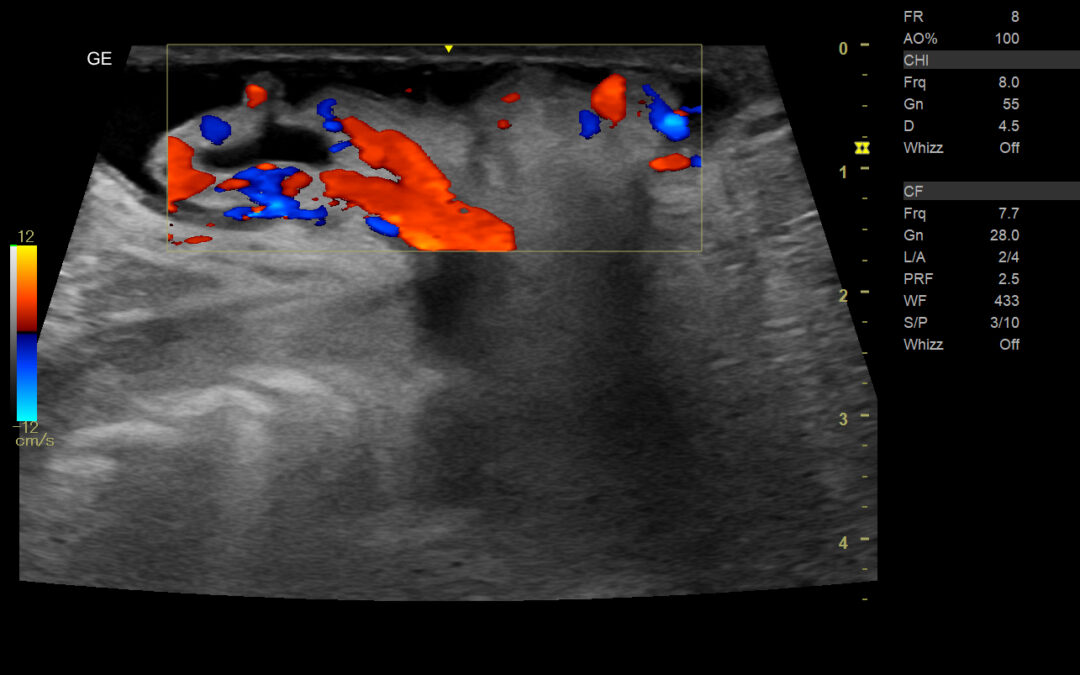
Images:


On color doppler evaluation there is evidence of mesenteric vessel showing significant vascularity along with the herniation of fat.
Ultrasound images shows an abdominal wall defect in the umbilical region with herniation omental and evidence free fluid in the herniated sac.
