by Nithesh Ravindran | Dec 13, 2021
Age: 36
Sex: Male
Complaints: C/o right parotid swelling.
Case study : Right parotid gland appears enlarged measures ~5.0 x 3.0 cm with an hypoechoic lesion showing mildly lobulated margins measuring ~3.5 x 2.0 cm with heteroechoic internal echotexture. Minimal internal vascularity. No evidence of calcification.
Images:
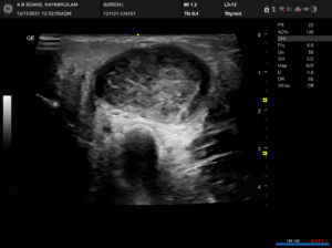
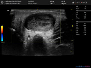
Conclusion: Well defined mildly lobulated solid hypoechoic lesion in the right parotid gland involving the superficial lobe as described. – Features in favor of Pleomorphic adenoma.
by Nithesh Ravindran | Dec 10, 2021
Name: Sudevan
Age: 54
Sex: Male
Complaints:
Introduction: H/o trauma
Case study: Thin linear hyperdensity noted involving the posterior inter-hemispheric fissure and right tentorium cerebelli. – Suggestive of acute subdural hemorrhage (SDH).
Images
Conclusion: Acute subdural hemorrhage involving posterior inter-hemispheric fissure and right tentorium cerebelli.
Reference :
by Nithesh Ravindran | Dec 10, 2021
Name: Bagini
Age: 78 Yrs
Sex: Female
Complaints: Lower abdominal pain
Introduction:
Case study: Both kidneys are seen joining at their lower poles and seen in the midline. The left kidney is seen partially in the renal fossa.
Images
Conclusion: Both kidneys are seen in the midline by joining their lower poles. – Horseshoe kidney.
Reference :
by Nithesh Ravindran | Dec 10, 2021
Age: 69 Yrs
Sex: Male
Complaints: Neck swelling.
Case study: An heterogenous appearing mass lesion measuring ~4.5 x 1.5 cm noted in the right postero-lateral aspect of neck involving the level III and VA with tiny calcifications and multiple irregular cystic lesions, largest measuring ~3.3 x 2.6 cm with internal echoes.
Images:
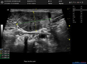
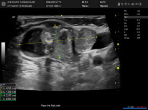
Conclusion: Solid heterogenous mass lesion in the right lateral aspect of neck involving level III and VA with cystic changes as described (TIRADS category 5). ? Metastatic lymphnode.
by Nithesh Ravindran | Dec 9, 2021
Name: Jaleela Ahammed
Age: 59 Yrs
Sex: Female
Complaints: To r/o abscess.
Introduction:
Case study: Well defined thick walled heterogenous appearing hypoechoic collection measuring ~24 x 12 mm noted in the subcutaneous region of left lower abdomen. No evidence of any internal vascularity.
Images
Conclusion: Well defined thick walled hypoechoic collection in the subcutaneous plane of left lower abdomen at the site of needle puncture. – Features in favour of post injection abscess.
Reference :