by Nithesh Ravindran | Dec 14, 2021
Name: Ramachandran
Age: 73 Yrs
Sex: Male
Complaints: H/o fall. Headache
Introduction:
Case study: Bilateral cerebral hemisphere shows well defined irregular hypodense (16-20HU) encapsulated subdural collection with maximum thickness measuring ~7 mm in the left high parietal region.
Images
Conclusion: Acute on chronic subdural hematoma (SDH) involving bilateral cerebral hemisphere as described.
Reference :
by Nithesh Ravindran | Dec 14, 2021
Case: 23
Heading: Acute on chronic SDH
Name: Dr. Ahammed Kunju
Age: 79 Yrs
Sex: Male
Complaints: H/o fall.
Introduction:
Case study: Bilateral cerebral hemisphere shows well defined encapsulated hypodense subdural collection with maximum thickness in the right measuring ~12 mm and seen extending along the parieto-occipital convexity showing irregular areas of hyperdensities.
Images
Conclusion: Well defined encapsulated hypodense subdural collection with randomly distributed areas of hyperdensity in the bilateral cerebral hemisphere. – Features suggestive of acute on chronic subdural hematoma.
Reference :
by Nithesh Ravindran | Dec 14, 2021
Age: 27 Yrs
Sex: Female
Complaints: H/o right breast pain
Introduction:
Case study: Well circumscribed irregular hypoechoic fluid collection measuring ~3.7 x 1.8 cm with complex mixed echogenicity showing peripheral vascularity and surrounding echogenic fat noted in the right upper midline at 12 o’clock position extending into the subareolar region.
Images:
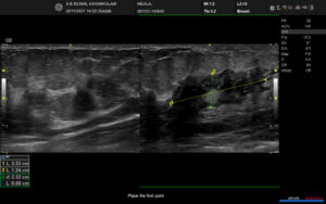

Conclusion: Well circumscribed irregular hypoechoic complex collection in the right breast extending into the subareolar region with surrounding inflammatory changes (BIRADS category 2). – Features in favour of acute mastitis with abscess formation.
Reference :
by Nithesh Ravindran | Dec 14, 2021
Name: Gowrikutty
Age: 80 Yrs
Sex: Female
Complaints:
Introduction:
Case study: Diffuse degenerative changes in the form of flowing osteophytes in the anterior aspect of thoracic vertebrae with loss of disc space and fusion of multiple vertebral bodies.
Images
Conclusion: Diffuse degenerative changes in the thoracic vertebrae in the form of flowing osteophytes, disc space reduction and fusion of multiple vertebrae bodies. – Features in favour of diffuse idiopathic skeletal hyperostosis (DISH).
Reference :
by Nithesh Ravindran | Dec 13, 2021
Name: Mukesh
Age: 32 Yrs
Sex: Male
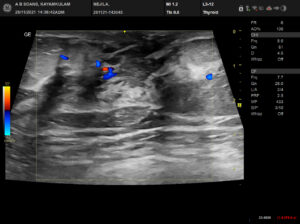
Complaints: C/o neck swelling
Introduction:
Case study: Well defined thick walled complex cystic lesion with minimal low level internal echoes measuring ~4.2 x 1.8 cm noted in the anterior midline of neck superior to thyroid gland. No significant internal vascularity. Peripheral vascularity noted.
Images
Conclusion: Well defined thick walled complex cystic lesion in the anterior midline of neck superior to normal thyroid gland. – Features suggestive of thyroglossal duct cyst
Reference :