by Nithesh Ravindran | Mar 15, 2023
Case courtesy : Dr.Cicy Kuncherian Welcare Hospital
Age: 56Yrs
Sex: Female
Complaints: Right iliac fossa pain.
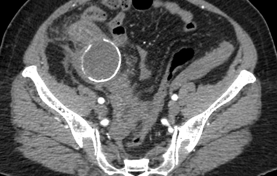
Case study: Tubular cystic mass lesion with peripheral enhancing walls measuring approximately 5.2 x4.3 x5.6 cm (TR XAP x CC) is seen in the right iliac fossa showing thick curvilinear mural calcification and central fluid density. No intraluminal air pockets noted.
The lesion is closely adherent towards the adjacent bowel loops causing bowel wall thickening of terminal ileum and cecum and adjacent fat stranding . Appendix not separately visualized from the mass lesion. Few prominent mesenteric lymph nodes are noted in right iliac fossa and paraumbilical region.
Small wall defect noted in the inferior aspect of the lesion with tubular extension measuring 14 x12mm extending into the right adnexa . Right ovary not separately
Images:
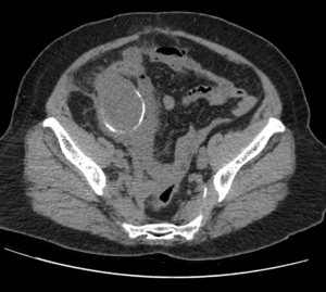
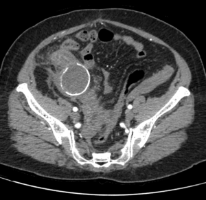
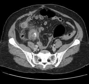
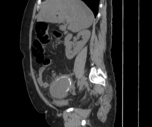
Conclusion : Tubular cystic mass lesion with curvilinear mural calcification in right iliac fossa with adjacent bowel wall thickening and fat stranding -features are in favor of Appendiceal Mucocele with contained perforation. Suggested lab correlation.
by Nithesh Ravindran | Mar 15, 2023
Case courtesy : Dr. Cicy Kuncheria Welcare Hospital
Age: 61Yrs
Sex: Female
Complaints: Recurrent pain over left mid-clavicular region.
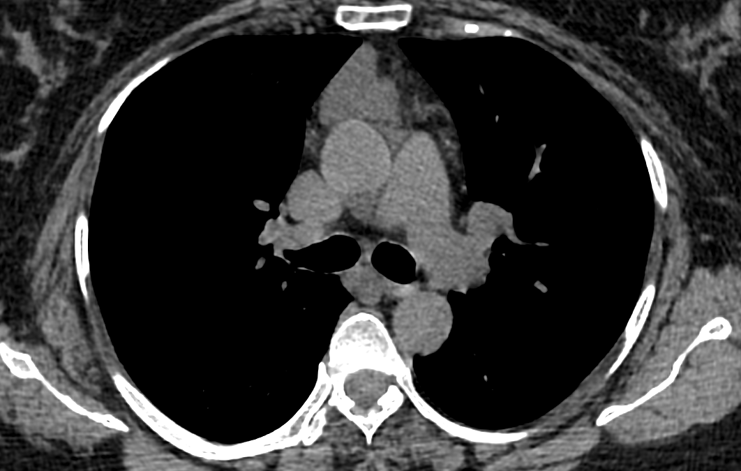
Case study: A smooth well defined soft tissue mass with fluid attenuation in the anterior mediastinum measuring ~3.9 x 2.9 x 3.4cm (TR XAP XCC).The lesion shows homogenous density.No calcification.
Images:
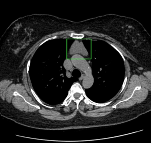
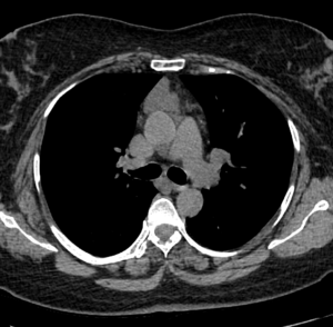
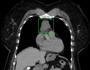
Conclusion : Smooth well defined anterior mediastinal mass lesion with fluid attenuation -imaging features are in favor of cystic mediastinal mass lesion. DD: Thymic cyst
by Nithesh Ravindran | Mar 15, 2023
Case courtesy : Dr.Cicy Kuncheria Welcare Hospital
Age: 34Yrs
Sex: Male
Complaints: Foot pain
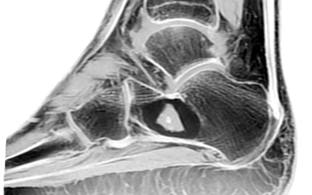
Case study: A calcaneal lesion with central hypointense signal on T1 and hyperintense on T2/PDFS with evidence of multiple blooming foci (calcifications on CT correlation).The lesion shows peripheral hyperintensity on T1 with PDsuppression.
Images:
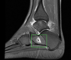
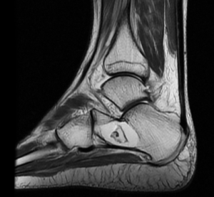

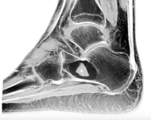
Conclusion : Sharply delineated lesion with predominant fat density. -Features in favour of an intraosseous lipoma of calcaneus – Stage 2.
by Nithesh Ravindran | Mar 15, 2023
Case courtesy : Dr.Cicy Kuncherian,Welcare Hospital.
Age: 27 Yrs
Sex: Male
Complaints: Swelling frontal region.
Case study: A well defined soft tissue density lesion (~ +13HU) measuring ~13 x 11 mm noted in the frontal region involving subgaleal layer. No evidence of intracranial invasion / bone erosion.
Images:

Conclusion :A well defined soft tissue lesion involving the mid frontal region causing superficial skin asymmetry as described – Imaging features in favour of dermoid cyst. Possible DD: Sebaceous cyst.

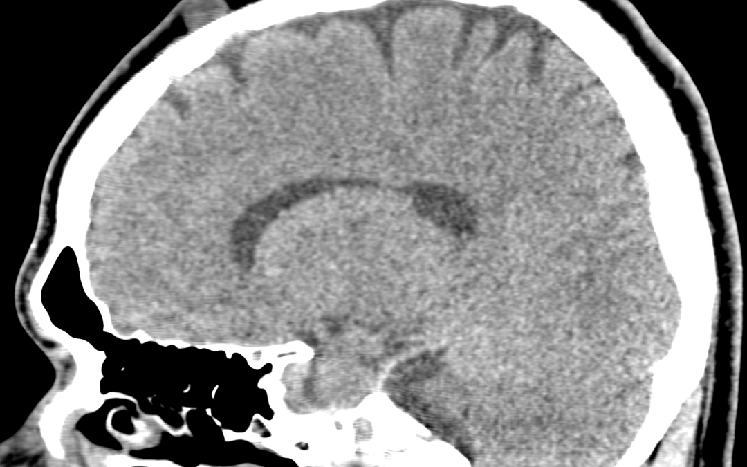
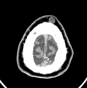
by Nithesh Ravindran | Dec 5, 2022
Case Courtesy : Dr Sajith Selvaganesan MD FSCCT FSCMR,Sunrise Hospital
Age: 25Yrs
Sex: Male
Complaints: Lower back pain.
Case study:
Loss of normal lumbar lordosis.
Relatively well defined, mildly heterogeneous, round to oval, iso to hyperintense intramedullary lesion measuring 11 x 10 x 17 mm (TR x AP x CC) noted the level of L3 vertebra
Images:
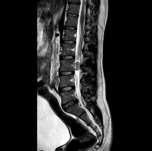
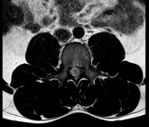
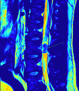
Conclusion
Relatively well defined, mildly heterogeneous, round to oval, iso to hyperintense intramedullary lesion measuring 11 x 10 x 17 mm (TR x AP x CC) noted the level of L3 vertebra – Likely myxopapillary ependymoma. Differential includes paraganglioma. Suggested HPE evaluation.