Head
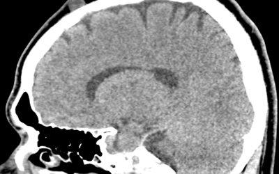
Scalp dermoid cyst
Case courtesy : Dr.Cicy Kuncherian,Welcare Hospital. Age: 27 Yrs Sex: Male Complaints: Swelling frontal region. Case study: A well defined soft tissue density lesion (~ +13HU) measuring ~13 x 11 mm noted in the frontal region involving subgaleal layer. No evidence of...
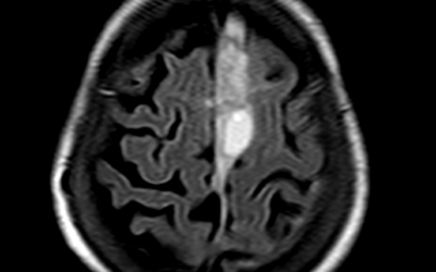
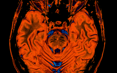
Subdural Hemorrhage MRI
Case courtesy : Dr. Sreedevi Varun DMRD DNB, Sunrise Hospital Age: 77Yrs Sex: Female Complaints: H/o fall. Case study: Area of heterogeneous T2 hyperintense signal in midline falx with maximum diameter 14 mm in highfrontal region. Cyst like area measuring 26 x 24 mm...
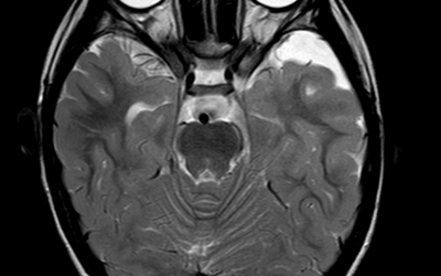
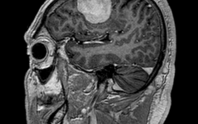
Arachnoid Cyst
Case courtesy : Dr.Sreedevi Varun DMRD DNB, Sunrise Hospital Age: 4Yrs Sex: Male Case study: An extra axial CSF intensity lesion in left anterior temporal fossa measuring 3.3 x 1.3 x 3.5 cm (TR x AP x CC) with no significant mass effect or perilesional...
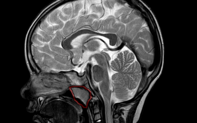
Adenoid Gland Hypertrophy
Age: 2Yrs Sex: Male Complaints: Speech Difficulty. Case study: Adenoid gland appears enlarged measuring ~ 16 .8mm (parasagittal). There is evident narrowing of nasopharyngeal air way. Images: Conclusion : Adenoid gland hypertrophy with nasopharyngeal airway...
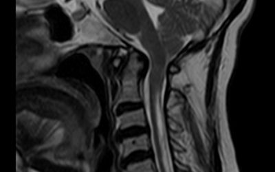
Chiari 1 Malformation with Syringohydromyelia
Case courtesy : Dr. Sameer Hyderali MD, Sunrise Hospital Age: 55Yrs Sex: Female Case study: Low placed cerebellar tonsils 5 mm below the foramen magnum. Tight foramen magnum with effaced 4th ventricle and the cisternal spaces noted. The cervico-medullary junction...
Meningioma
Age: 65Yrs Sex: Male Complaints: Head ache Case study: Moderately enhancing right posterior parietal foci showing broad base to the dura measuring 1.8 x 3.4 x 2.5 cm. The foci shows evidence of dural tail with evidence of indentation on the adjacent brain parenchyma...
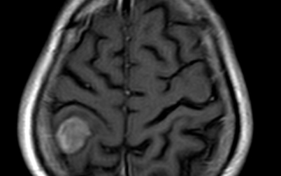
Typical Meningioma
Age: 59yrs Sex: Male Complaints: Case study: Supratentorial extra-axial dural based well defined lesion in right frontal convexity measuring 4.2 x 2.6 x 3.5 cm (AP x TR x CC) causing moderate perilesional edema and mild mass effect on adjacent brain parenchyma seen....
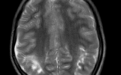
Hypoxic Ischemic Encephalopathy (HIE)
Age: 11 Yrs Sex: Female Complaints: Developmental abnormalities. K/c/o HIE. Case study: Evidence of periventricular T2/FLAIR hyperintensities seen in the posterior fossa around the occipital horn extending into the adjacent sulcal spaces. The corpus callosum...
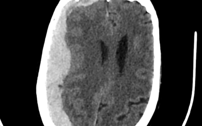
Spontaneous Non-Traumatic SDH
Age: 48yr Sex: Female Complaints: Sudden headache and unconsciousness. Case study: Extra axial, crescent shaped, hyperdense areas noted along the right temporo-fronto-parietal convexity (maximum thickness ~22 mm) causing mass effect in the form of effacement of...
Autosomal Recessive Spastic Ataxia of Charlevoix-Saguenay
Age: 28yr Sex: Male Complaints: Progressive walking difficulty. Case study: T2 hypointensity noted in the pons and midbrain region with mild reduction in the bulk of the vermis. Mild prominence of the cisterna magna noted. The cerebellar foliae appears mildly...